


Organs as
Never Seen Before
The field of medical imaging has come a long way since 1895, when German physicist Wilhelm Röntgen aimed an electron beam at his wife's hand and imaged her bones and wedding ring on a photographic plate, generating what's believed to be the first X-ray. Today, millions of radiological images are created every year in the U.S., thanks to a growing suite of sophisticated tools, several of which have been advanced by researchers at NYU Langone.
- The Chandarana Lab
- The Fishman Lab
- The He Lab
- The Chandarana Lab
- The Fishman Lab
- The He Lab
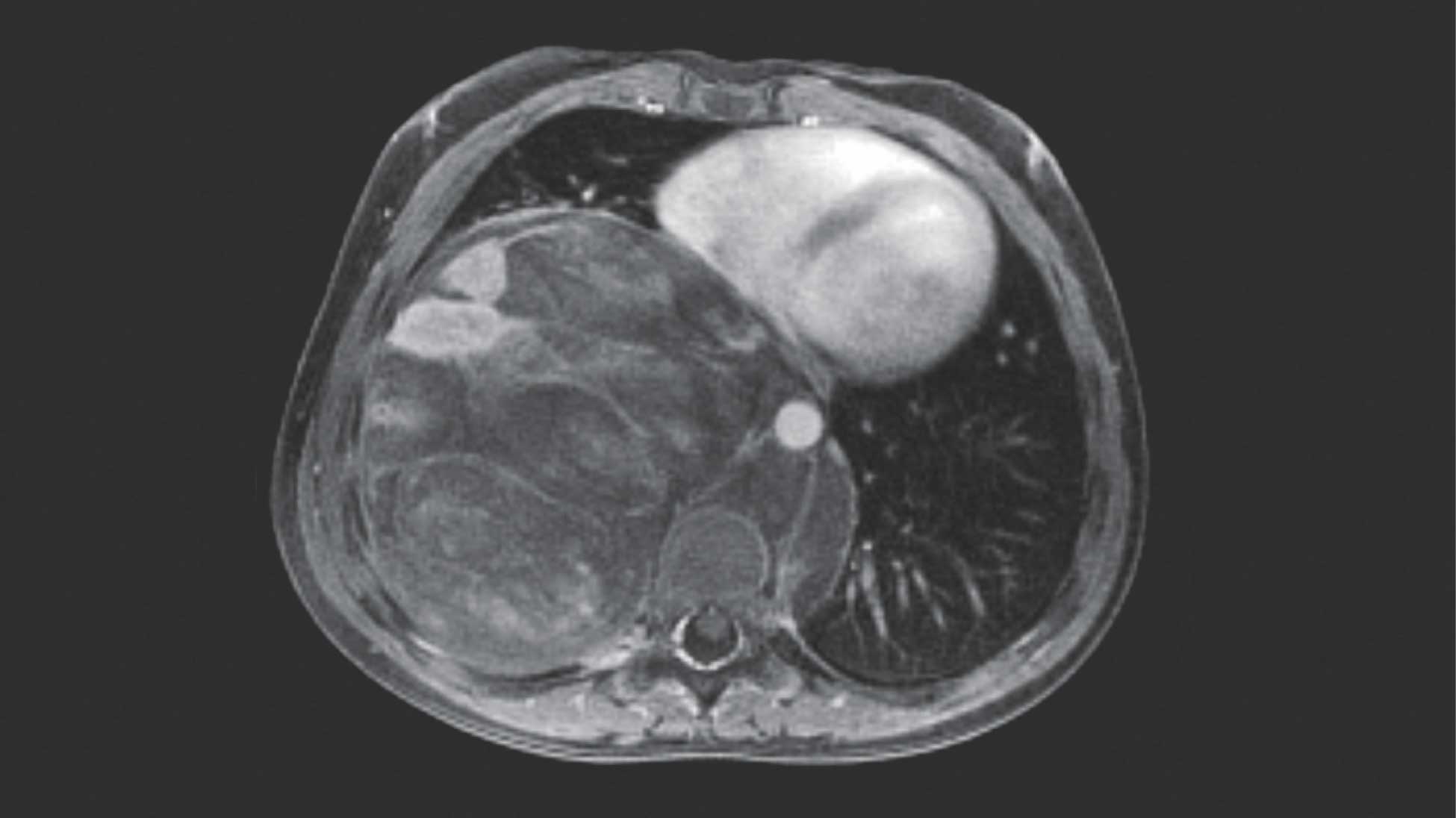
With a conventional MRI, the borders of the large pale mass shown at right would be indistinct in this six-year-old patient.
Dr. Chandarana's free-breathing-motion robust MRI shows much crisper borders and confirms that the mass is contacting but
not invading the patient's heart.
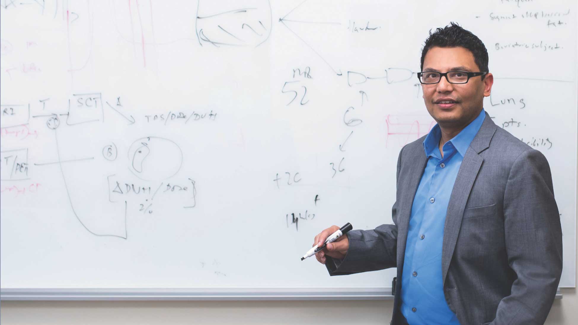
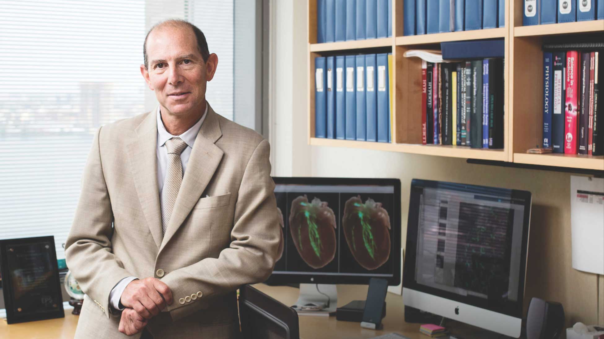
Hersh Chandarana, MD
Associate professor of radiology and section chief of the Division of Body Imaging at NYU Langone
Associate professor of radiology and section chief of the Division of Body Imaging at NYU Langone
1 of 2
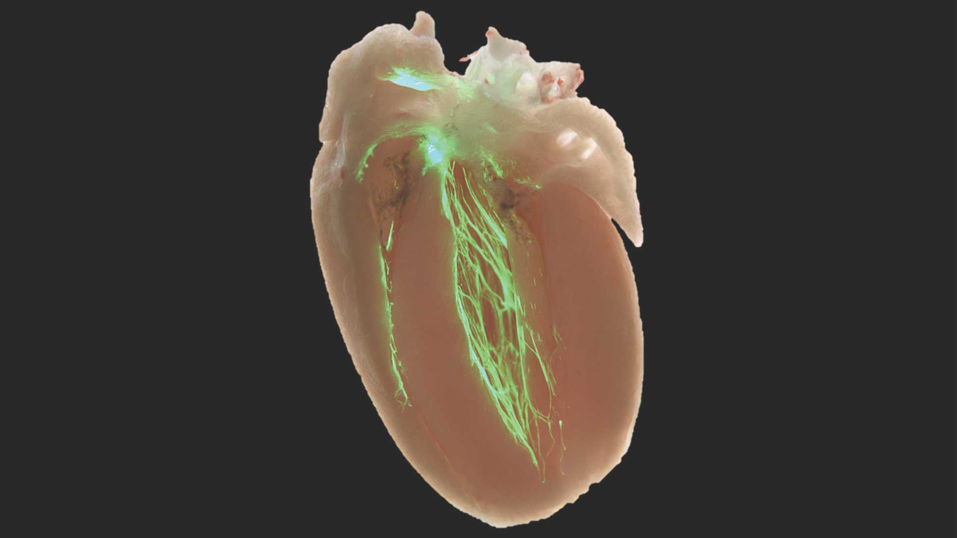
Specialized heart cells called Purkinje cells are shown here within a mouse heart as a fluorescent green treelike
network that forms the cardiac conduction system.
Glenn Fishman, MD
William Goldring professor of medicine and director of the Leon H. Charney Division of Cardiology at NYU Langone
William Goldring professor of medicine and director of the Leon H. Charney Division of Cardiology at NYU Langone
1 of 2
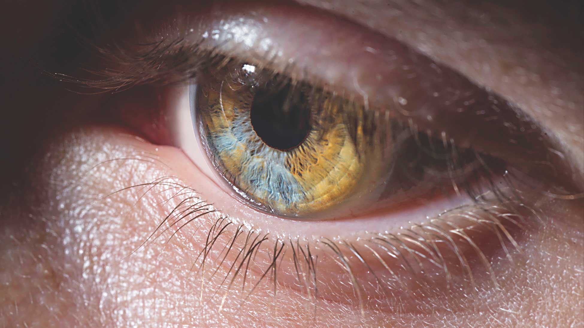
When we perceive our surroundings, that conscious awareness is generated by the brain through an unknown process. The He lab
is using cutting-edge imaging techniques and studying brain activity patterns in human volunteers to better understand how
varying brain activity gives rise to different conscious experiences.
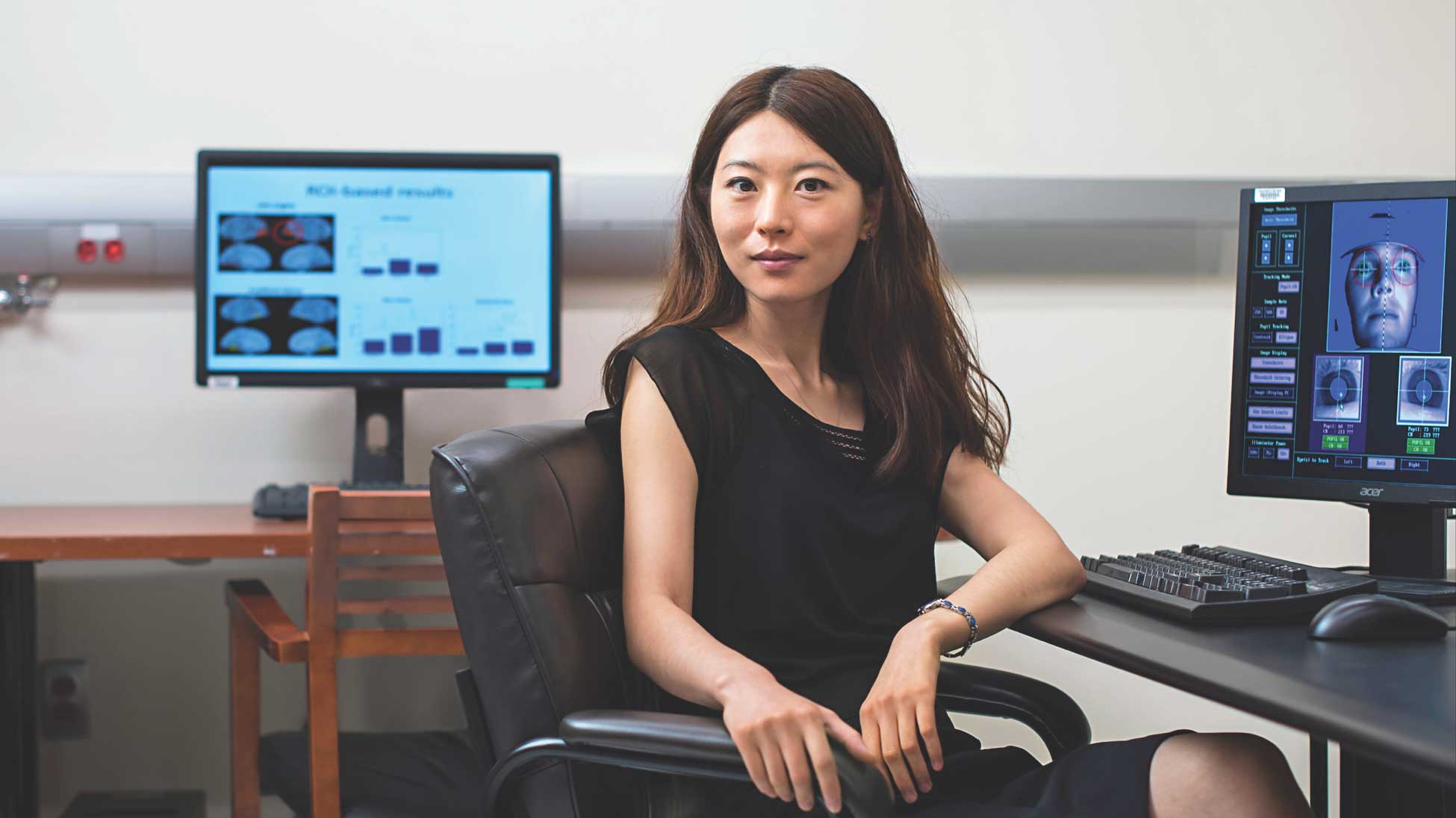
Biyu Jade He, PhD
Assistant professor of neurology, neuroscience and physiology, and radiology at NYU Langone
Assistant professor of neurology, neuroscience and physiology, and radiology at NYU Langone
1 of 2