


Cells in Their
Natural Habitats
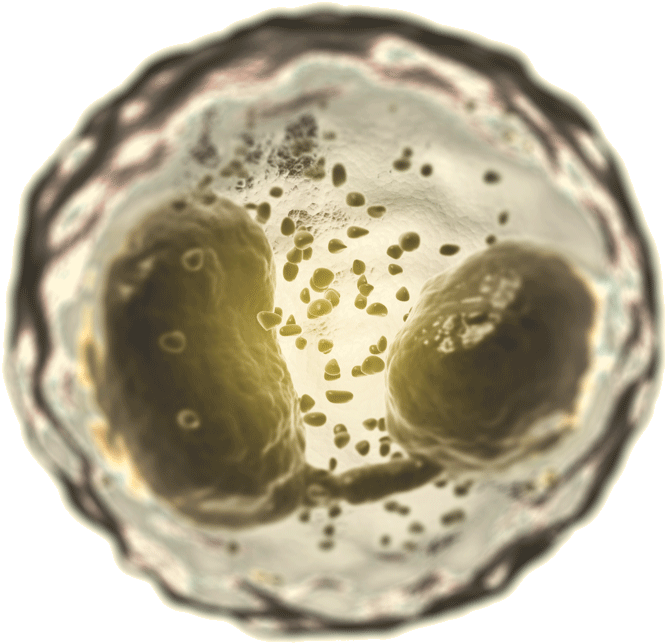
In 1653, a 26-year-old self-educated prodigy named Robert C. Hooke was studying a thin slice of cork through the lens
of a homemade microscope when he was struck by an unusual pattern of compartments that he likened to misshapen honeycomb.
He dubbed the structures cells,
and scientists have been using the term ever since to describe the basic building blocks
of life. Today, remarkable advances in microscopy afford researchers a view of these building blocks that Hooke could
scarcely have imagined. At NYU Langone, such advances are helping biologists understand how a dizzying number of interactions
among the body's 30 trillion cells can spark disease.
- The Schwab Lab
- The Rinberg Lab
- The Schwab Lab
- The Rinberg Lab
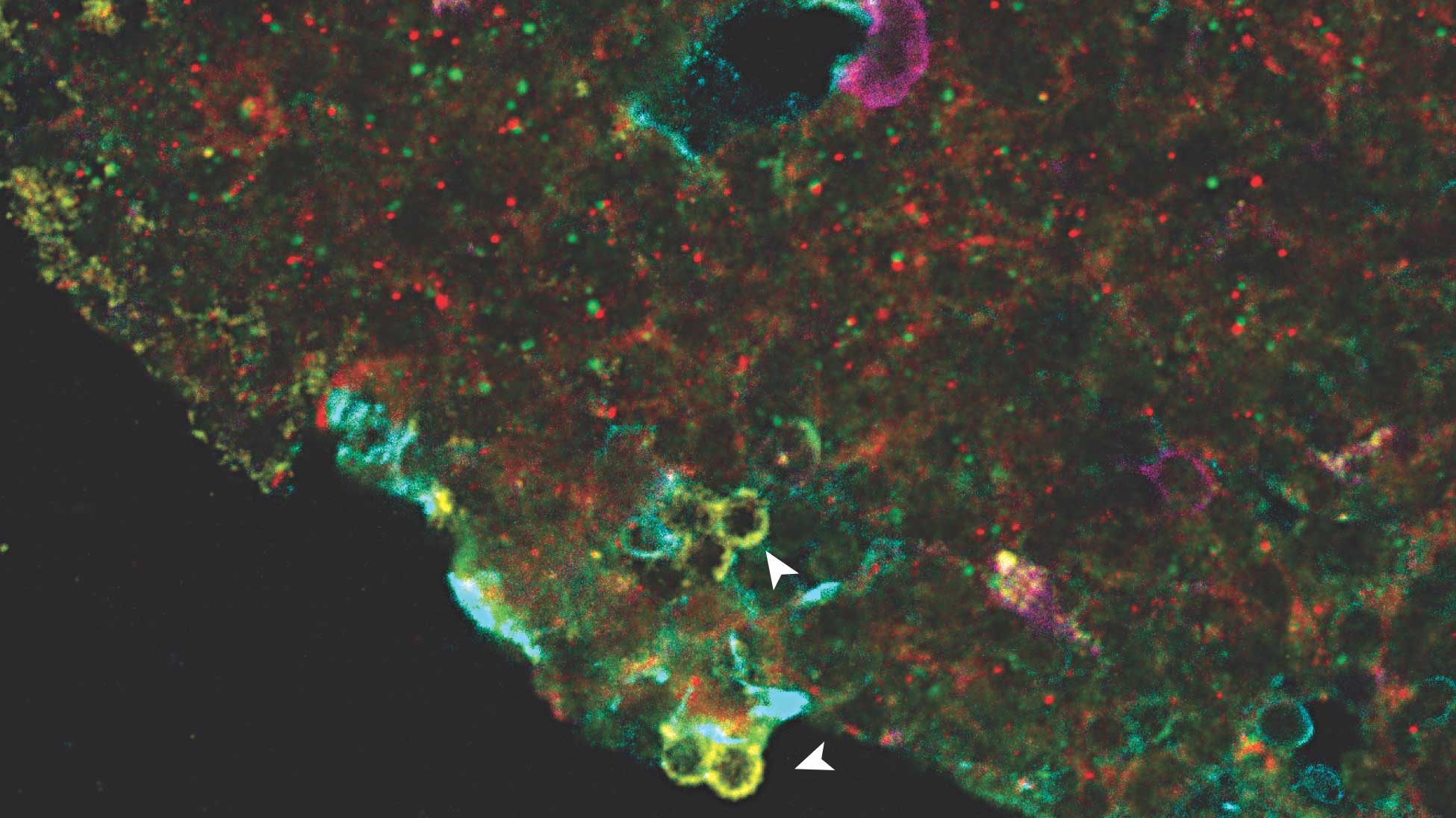
This image of mouse lymph node cells shows green and red fluorescent labels used by the Schwab lab to tag a surface
protein that normally binds to the S1P molecule. Tiny green dots indicate cells where the surface protein has
recognized and bound to S1P, while the red label indicates a defective protein variant that no longer recognizes
the molecule. A yellowish hue suggests the cell hasn’t encountered S1P at all.
Susan Schwab, PhD
Assistant professor of pathology at the Skirball Institute of Biomolecular Medicine at NYU Langone
Assistant professor of pathology at the Skirball Institute of Biomolecular Medicine at NYU Langone
1 of 2

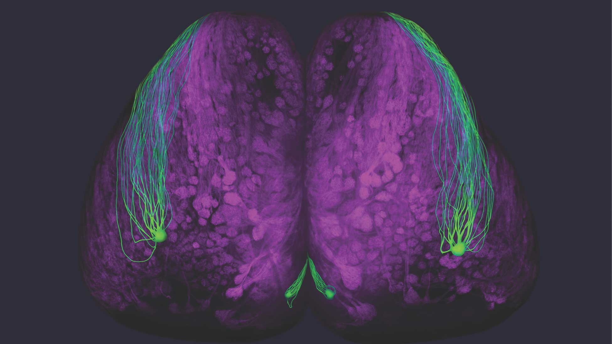
The Rinberg lab uses a variety of odors (shown in bottles) to understand the neural basis of olfaction in mice.
This illuminated image of a mouse brain demonstrates a light-based technique called optogenetics, in which the lab uses a laser to
turn onspecific sensory neurons (represented by green dots) that initiate the brain’s odor recognition pathway.
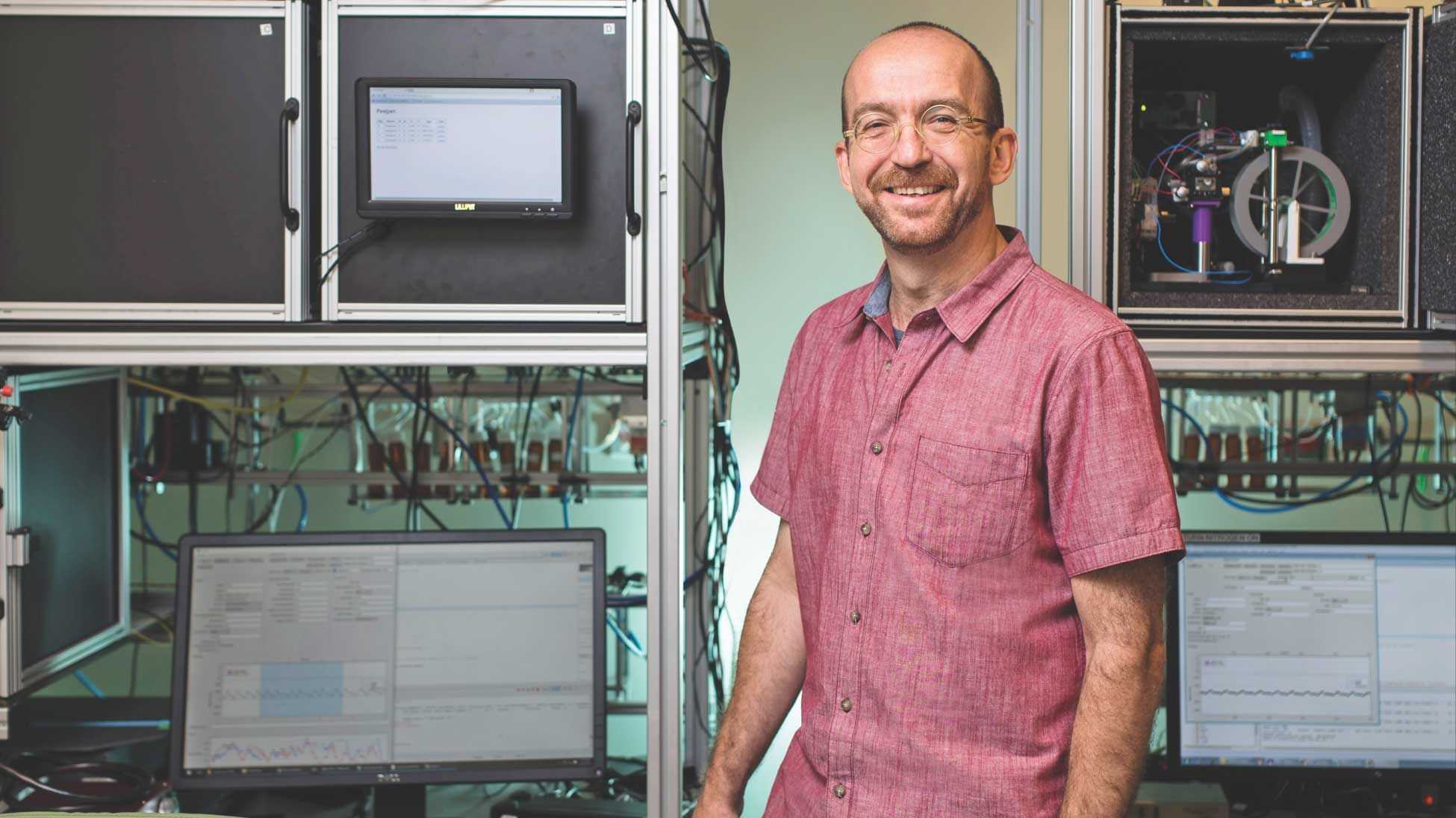
Dmitry Rinberg, PhD
Associate professor of neuroscience and physiology at NYU Langone’s Neuroscience Institute
Associate professor of neuroscience and physiology at NYU Langone’s Neuroscience Institute
1 of 3