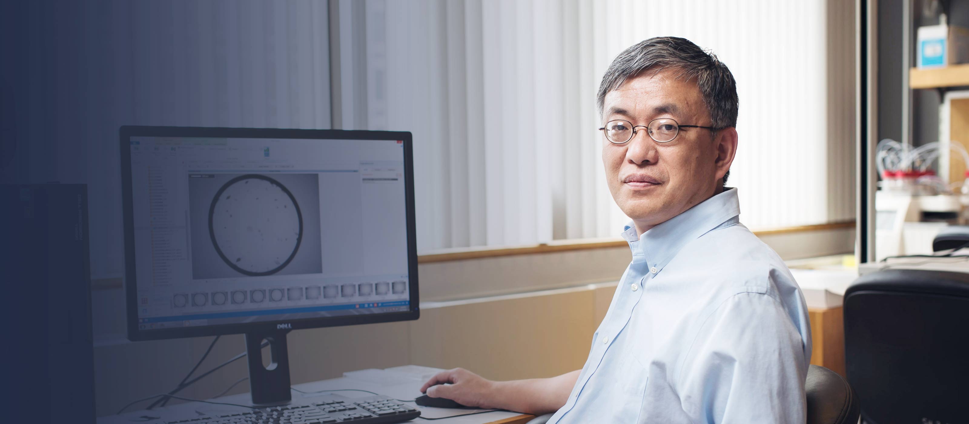
Da-Neng Wang

Biosketch / Results /

Da-Neng Wang, Ph.D.

Professor;
Department of Cell Biology (Skirball) and Structural Biology

Contact Info

Address
540 First Avenue
Floor 3 Room 3-Lab 5
Skirball Institute
New York, NY 10016

212-263-8634
212-263-8951
Da-Neng.Wang@med.nyu.edu


« Back to Results

Education

1988 — University of Stockholm, Graduate Education
— Dr. Wang was a Postdoctoral Fellow at the European Molecular Biology Laboratory (EMBL), Heidelberg, Germany, PostDoctoral Training

« Back to Results

Research Interests

Structural Biology of Membrane Proteins<br>

Research Keywords

cryo-electron microscopy, X-ray crystallography, membrane protein structure, transporters<br>

« Back to Results

All data from NYU Health Sciences Library Faculty Bibliography — -

Contact:
http://hsl.med.nyu.edu/faculty-bibliography-search#about

Simple screening method for improving membrane protein thermostability
Mancusso, Romina; Karpowich, Nathan K; Czyzewski, Bryan K; Wang, Da-Neng
2011 Dec;55(4):324-329, Methods
Biochemical and biophysical analysis on integral membrane proteins often requires monodisperse and stable protein samples. Here we describe a method to characterize protein thermostability by measuring its melting temperature in detergent using analytical size-exclusion chromatography. This quantitative method can be used to screen for compounds and conditions that stabilize the protein. With this technique we were able to assess and improve the thermostability of several membrane proteins. These conditions were in turn used to assist purification, to identify protein ligand and to improve crystal quality
— id: 150557, year: 2011, vol: 55, page: 324, stat: Journal Article,

GEP constitutes a negative feedback loop with MyoD and acts as a novel mediator in controlling skeletal muscle differentiation
Wang D; Bai X; Tian Q; Lai Y; Lin EA; Shi Y; Mu X; Feng JQ; Carlson CS; Liu CJ
2011 Dec 17;:?-? #, Cellular & molecular life sciences: CMLS
Granulin-epithelin precursor (GEP) is an autocrine growth factor that has been implicated in embryonic development, tissue repair, tumorigenesis, and inflammation. Here we report that GEP was expressed in skeletal muscle tissue and its level was differentially altered in the course of C2C12 myoblast fusion. The GEP expression during myoblast fusion was a consequence of MyoD transcription factor binding to several E-box (CANNTG) sequences in the 5'-flanking regulatory region of GEP gene, followed by transcription. Recombinant GEP potently inhibited myotube formation from C2C12 myoblasts whereas the knockdown of endogenous of GEP via a siRNA approach accelerated the fusion of myoblasts to myotubes. Interestingly, the muscle fibers of GEP knockdown mice were larger in number but noticeably smaller in size when compared to the wild-type. Mechanistic studies revealed that during myoblast fusion, the addition of GEP led to remarkable reductions in the expressions of muscle-specific transcription factors, including MyoD. In addition, the regulation of myotube formation by GEP is mediated by the anti-myogenic factor JunB, which is upregulated following GEP stimulation. Thus, GEP growth factor, JunB, and MyoD transcription factor form a regulatory loop and act in concert in the course of myogenesis
— id: 148767, year: 2011, vol: , page: ?, stat: Journal Article,

Protein Signatures for Classification and Prognosis of Gastric Cancer A Signaling Pathway-Based Approach
Wang, Daguang; Ye, Fei; Sun, Yabin; Li, Wei; Liu, Hongyi; Jiang, Jing; Zhang, Yang; Liu, Chengkui; Tong, Weihua; Gao, Ling; Sun, Yezhou; Zhang, Weijia; SeeToe, Terry; Lee, Peng; Suo, Jian; Zhang, David Y.
2011 OCT ;179(4):1657-1666, American journal of pathology
Current methods have limited accuracy in predicting survival and stratifying patients with gastric cancer for appropriate treatment. We sought to identify protein signatures of gastric cancer for classification and prognostication. The Protein Pathway Array (initial study) and Western blot (confirmation) were used to assess the protein expression in a total of 199 fresh frozen gastric samples. There were 56 paired samples divided into a training set (n = 37) and a validation set (n = 19) for the identification of differentially expressed proteins between tumor and normal tissues. There were 56 tumor samples used to identify proteins correlating with tumor and nodal staging. All 93 tumor samples were used to identify candidate proteins for predicting survival. We confirmed the survival prediction of the candidate proteins by using an additional cohort of gastric cancer samples (n = 50). There were 22 proteins differentially expressed between normal and tumor tissues. Nine proteins were selected to build the predictor to classify normal and tumor samples. Ten proteins were differentially expressed among different T stages and four of these were associated with invasive behavior. An additional four proteins were associated with lymph node metastasis. Two proteins were identified as independent risk factors for overall survival. This study indicated that some dysregulated signaling proteins could be selected as useful biomarkers for tumor classification and predicting outcome in gastric cancer patients. (Am J Pathol 2011, 179:1657-1666; DOI: 10.1016/j.ajpath.2011.06.010)
— id: 149889, year: 2011, vol: 179, page: 1657, stat: Journal Article,

Biophysics: Transporter in the spotlight
Karpowich, Nathan K; Wang, Da-Neng
2010 May 13;465(7295):171-172, Nature
— id: 132714, year: 2010, vol: 465, page: 171, stat: Journal Article,

The New York Consortium on Membrane Protein Structure (NYCOMPS): a high-throughput platform for structural genomics of integral membrane proteins
Love, James; Mancia, Filippo; Shapiro, Lawrence; Punta, Marco; Rost, Burkhard; Girvin, Mark; Wang, Da-Neng; Zhou, Ming; Hunt, John F; Szyperski, Thomas; Gouaux, Eric; MacKinnon, Roderick; McDermott, Ann; Honig, Barry; Inouye, Masayori; Montelione, Gaetano; Hendrickson, Wayne A
2010 Sep;11(3):191-199, Journal of structural & functional genomics
The New York Consortium on Membrane Protein Structure (NYCOMPS) was formed to accelerate the acquisition of structural information on membrane proteins by applying a structural genomics approach. NYCOMPS comprises a bioinformatics group, a centralized facility operating a high-throughput cloning and screening pipeline, a set of associated wet labs that perform high-level protein production and structure determination by x-ray crystallography and NMR, and a set of investigators focused on methods development. In the first three years of operation, the NYCOMPS pipeline has so far produced and screened 7,250 expression constructs for 8,045 target proteins. Approximately 600 of these verified targets were scaled up to levels required for structural studies, so far yielding 24 membrane protein crystals. Here we describe the overall structure of NYCOMPS and provide details on the high-throughput pipeline
— id: 132716, year: 2010, vol: 11, page: 191, stat: Journal Article,

Substrate and drug binding sites in LeuT
Nyola, Ajeeta; Karpowich, Nathan K; Zhen, Juan; Marden, Jennifer; Reith, Maarten E; Wang, Da-Neng
2010 Aug;20(4):415-422, Current opinion in structural biology
LeuT is a member of the neurotransmitter/sodium symporter family, which includes the neuronal transporters for serotonin, norepinephrine, and dopamine. The original crystal structure of LeuT shows a primary leucine-binding site at the center of the protein. LeuT is inhibited by different classes of antidepressants that act as potent inhibitors of the serotonin transporter. The newly determined crystal structures of LeuT-antidepressant complexes provide opportunities to probe drug binding in the serotonin transporter, of which the exact position remains controversial. Structure of a LeuT-tryptophan complex shows an overlapping binding site with the primary substrate site. A secondary substrate binding site was recently identified, where the binding of a leucine triggers the cytoplasmic release of the primary substrate. This two binding site model presents opportunities for a better understanding of drug binding and the mechanism of inhibition for mammalian transporters
— id: 112042, year: 2010, vol: 20, page: 415, stat: Journal Article,

Structure and mechanism of a pentameric formate channel
Waight, Andrew B; Love, James; Wang, Da-Neng
2010 Jan;17(1):31-37, Nature structural & molecular biology
Formate transport across the inner membrane is a critical step in anaerobic bacterial respiration. Members of the formate/nitrite transport protein family function to shuttle substrate across the cytoplasmic membrane. In bacterial pathogens, the nitrite transport protein is involved in protecting bacteria from peroxynitrite released by host macrophages. We have determined the 2.13-A structure of the formate channel FocA from Vibrio cholerae, which reveals a pentamer in which each monomer possesses its own substrate translocation pore. Unexpectedly, the fold of the FocA monomer resembles that found in water and glycerol channels. The selectivity filter in FocA consists of a cytoplasmic slit and a central constriction ring. A 2.5-A high-formate structure shows two formate ions bound to the cytoplasmic slit via both hydrogen bonding and van der Waals interactions, providing a structural basis for the substrate selectivity of the channel
— id: 106095, year: 2010, vol: 17, page: 31, stat: Journal Article,

Structural basis of substrate selectivity in the glycerol-3-phosphate: phosphate antiporter GlpT
Law, Christopher J; Enkavi, Giray; Wang, Da-Neng; Tajkhorshid, Emad
2009 Sep 2;97(5):1346-1353, Biophysical journal
Major facilitators represent the largest superfamily of secondary active transporter proteins and catalyze the transport of an enormous variety of small solute molecules across biological membranes. However, individual superfamily members, although they may be architecturally similar, exhibit strict specificity toward the substrates they transport. The structural basis of this specificity is poorly understood. A member of the major facilitator superfamily is the glycerol-3-phosphate (G3P) transporter (GlpT) from the Escherichia coli inner membrane. GlpT is an antiporter that transports G3P into the cell in exchange for inorganic phosphate (P(i)). By combining large-scale molecular-dynamics simulations, mutagenesis, substrate-binding affinity, and transport activity assays on GlpT, we were able to identify key amino acid residues that confer substrate specificity upon this protein. Our studies suggest that only a few amino acid residues that line the transporter lumen act as specificity determinants. Whereas R45, K80, H165, and, to a lesser extent Y38, Y42, and Y76 contribute to recognition of both free P(i) and the phosphate moiety of G3P, the residues N162, Y266, and Y393 function in recognition of only the glycerol moiety of G3P. It is the latter interactions that give the transporter a higher affinity to G3P over P(i)
— id: 132717, year: 2009, vol: 97, page: 1346, stat: Journal Article,

STRUCTURE OF THE LEUCINE TRANSPORTER LeuT IN COMPLEX WITH DESIPRAMINE SUGGESTS HOW ANTIDEPRESSANTS INHIBIT NEUROTRANSMITTER REUPTAKE
Wang, DN
2009 FEB ;59(2):116-116, Journal of physiological sciences : JPS
— id: 106962, year: 2009, vol: 59, page: 116, stat: Journal Article,

Antidepressant specificity of serotonin transporter suggested by three LeuT-SSRI structures
Zhou, Zheng; Zhen, Juan; Karpowich, Nathan K; Law, Christopher J; Reith, Maarten E A; Wang, Da-Neng
2009 Jun;16(6):652-657, Nature structural & molecular biology
Sertraline and fluoxetine are selective serotonin re-uptake inhibitors (SSRIs) that are widely prescribed to treat depression. They exert their effects by inhibiting the presynaptic plasma membrane serotonin transporter (SERT). All SSRIs possess halogen atoms at specific positions, which are key determinants for the drugs' specificity for SERT. For the SERT protein, however, the structural basis of its specificity for SSRIs is poorly understood. Here we report the crystal structures of LeuT, a bacterial SERT homolog, in complex with sertraline, R-fluoxetine or S-fluoxetine. The SSRI halogens all bind to exactly the same pocket within LeuT. Mutation at this halogen-binding pocket (HBP) in SERT markedly reduces the transporter's affinity for SSRIs but not for tricyclic antidepressants. Conversely, when the only nonconserved HBP residue in both norepinephrine and dopamine transporters is mutated into that found in SERT, their affinities for all the three SSRIs increase uniformly. Thus, the specificity of SERT for SSRIs is dependent largely on interaction of the drug halogens with the protein's HBP
— id: 103150, year: 2009, vol: 16, page: 652, stat: Journal Article,

Three-dimensional Architecture of Hair-bundle Linkages Revealed by Electron-microscopic Tomography
Auer, Manfred; Koster, Abrahram J; Ziese, Ulrike; Bajaj, Chandrajit; Volkmann, Niels; Wang, Da Neng; Hudspeth, A J
2008 Jun;9(2):215-224, Journal of the Association for Research in Otolaryngology : JARO
The senses of hearing and balance rest upon mechanoelectrical transduction by the hair bundles of hair cells in the inner ear. Located at the apical cellular surface, each hair bundle comprises several tens of stereocilia and a single kinocilium that are interconnected by extracellular proteinaceous links. Using electron-microscopic tomography of bullfrog saccular sensory epithelia, we examined the three-dimensional structures of basal links, kinociliary links, and tip links. We observed significant differences in the appearances and dimensions of these three structures and found two distinct populations of tip links suggestive of the involvement of different proteins, splice variants, or protein-protein interactions. We noted auxiliary links connecting the upper portions of tip links to the taller stereocilia. Tip links and auxiliary links show a tendency to adopt a globular conformation when disconnected from the membrane surface
— id: 78730, year: 2008, vol: 9, page: 215, stat: Journal Article,

Structural biology. Symmetric transporters for asymmetric transport
Karpowich, Nathan K; Wang, Da-Neng
2008 Aug 8;321(5890):781-782, Science
— id: 81068, year: 2008, vol: 321, page: 781, stat: Journal Article,

Salt-bridge dynamics control substrate-induced conformational change in the membrane transporter GlpT
Law, Christopher J; Almqvist, Jonas; Bernstein, Adam; Goetz, Regina M; Huang, Yafei; Soudant, Celine; Laaksonen, Aatto; Hovmoller, Sven; Wang, Da-Neng
2008 May 9;378(4):826-837, Journal of molecular biology
Active transport of substrates across cytoplasmic membranes is of great physiological, medical and pharmaceutical importance. The glycerol-3-phosphate (G3P) transporter (GlpT) of the E. coli inner membrane is a secondary active antiporter from the ubiquitous major facilitator superfamily that couples the import of G3P to the efflux of inorganic phosphate (P(i)) down its concentration gradient. Integrating information from a novel combination of structural, molecular dynamics simulations and biochemical studies, we identify the residues involved directly in binding of substrate to the inward-facing conformation of GlpT, thus defining the structural basis for the substrate-specificity of this transporter. The substrate binding mechanism involves protonation of a histidine residue at the binding site. Furthermore, our data suggest that the formation and breaking of inter- and intradomain salt bridges control the conformational change of the transporter that accompanies substrate translocation across the membrane. The mechanism we propose may be a paradigm for organophosphate:phosphate antiporters
— id: 78697, year: 2008, vol: 378, page: 826, stat: Journal Article,

Ins and outs of major facilitator superfamily antiporters
Law, Christopher J; Maloney, Peter C; Wang, Da-Neng
2008 ;62:289-305, Annual review of microbiology
The major facilitator superfamily (MFS) represents the largest group of secondary active membrane transporters, and its members transport a diverse range of substrates. Recent work shows that MFS antiporters, and perhaps all members of the MFS, share the same three-dimensional structure, consisting of two domains that surround a substrate translocation pore. The advent of crystal structures of three MFS antiporters sheds light on their fundamental mechanism; they operate via a single binding site, alternating-access mechanism that involves a rocker-switch type movement of the two halves of the protein. In the sn-glycerol-3-phosphate transporter (GlpT) from Escherichia coli, the substrate-binding site is formed by several charged residues and a histidine that can be protonated. Salt-bridge formation and breakage are involved in the conformational changes of the protein during transport. In this review, we attempt to give an account of a set of mechanistic principles that characterize all MFS antiporters
— id: 93312, year: 2008, vol: 62, page: 289, stat: Journal Article,

Docking and homology modeling explain inhibition of the human vesicular glutamate transporters
Almqvist, Jonas; Huang, Yafei; Laaksonen, Aatto; Wang, Da-Neng; Hovmoller, Sven
2007 Sep;16(9):1819-1829, Protein science
As membrane transporter proteins, VGLUT1-3 mediate the uptake of glutamate into synaptic vesicles at presynaptic nerve terminals of excitatory neural cells. This function is crucial for exocytosis and the role of glutamate as the major excitatory neurotransmitter in the central nervous system. The three transporters, sharing 76% amino acid sequence identity in humans, are highly homologous but differ in regional expression in the brain. Although little is known regarding their three-dimensional structures, hydropathy analysis on these proteins predicts 12 transmembrane segments connected by loops, a topology similar to other members in the major facilitator superfamily, where VGLUT1-3 have been phylogenetically classified. In this work, we present a three-dimensional model for the human VGLUT1 protein based on its distant bacterial homolog in the same superfamily, the glycerol-3-phosphate transporter from Escherichia coli. This structural model, stable during molecular dynamics simulations in phospholipid bilayers solvated by water, reveals amino acid residues that face its pore and are likely to affect substrate translocation. Docking of VGLUT1 substrates to this pore localizes two different binding sites, to which inhibitors also bind with an overall trend in binding affinity that is in agreement with previously published experimental data
— id: 132718, year: 2007, vol: 16, page: 1819, stat: Journal Article,

Kinetic Evidence Is Consistent with the Rocker-Switch Mechanism of Membrane Transport by GlpT
Law, Christopher J; Yang, Qiang; Soudant, Celine; Maloney, Peter C; Wang, Da-Neng
2007 Oct 30;46(43):12190-12197, Biochemistry
Secondary active transport of substrate across the cell membrane is crucial to many cellular and physiological processes. The crystal structure of one member of the secondary active transporter family, the sn-glycerol-3-phosphate (G3P) transporter (GlpT) of the inner membrane of Escherichia coli, suggests a mechanism for substrate translocation across the membrane that involves a rocker-switch-type movement of the protein. This rocker-switch mechanism makes two specific predictions with respect to kinetic behavior: the transport rate increases with the temperature, whereas the binding affinity of the transporter to a substrate is temperature-independent. In this work, we directly tested these two predictions by transport kinetics and substrate-binding experiments, integrating the data on this single system into a coherent set of observations. The transport kinetics of the physiologically relevant G3P-phosphate antiport reaction were characterized at different temperatures using both E. coli whole cells and GlpT reconstituted into proteoliposomes. Substrate-binding affinity of the transporter was measured using tryptophan fluorescence quenching in detergent solution. Indeed, the substrate transport velocity of GlpT increased dramatically with temperature. In contrast, neither the apparent Michaelis constant (Km) nor the apparent substrate-binding dissociation constant (Kd) showed temperature dependence. Moreover, GlpT-catalyzed G3P translocation exhibited a completely linear Arrhenius function with an activation energy of 35.2 kJ mol-1 for the transporter reconstituted into proteoliposomes, suggesting that the substrate-loaded transporter is delicately poised between the inward- and outward-facing conformations. When these results are taken together, they are in agreement with a rocker-switch mechanism for GlpT
— id: 74677, year: 2007, vol: 46, page: 12190, stat: Journal Article,

LeuT-Desipramine Structure Reveals How Antidepressants Block Neurotransmitter Reuptake
Zhou, Zheng; Zhen, Juan; Karpowich, Nathan K; Goetz, Regina M; Law, Christopher J; Reith, Maarten E A; Wang, Da-Neng
2007 Sep 7;317(5843):1390-1393, Science
Tricyclic antidepressants exert their pharmacological effect-inhibiting the reuptake of serotonin, norepinephrine, and dopamine-by directly blocking neurotransmitter transporters (SERT, NET, and DAT, respectively) in the presynaptic membrane. The drug-binding site and the mechanism of this inhibition are poorly understood. We determined the crystal structure at 2.9 A of the bacterial leucine transporter (LeuT), a homolog of SERT, NET, and DAT, in complex with leucine and the antidepressant desipramine. Desipramine binds at the inner end of the extracellular cavity of the transporter and is held in place by a hairpin loop and by a salt bridge. This binding site is separated from the leucine-binding site by the extracellular gate of the transporter. By directly locking the gate, desipramine prevents conformational changes and blocks substrate transport. Mutagenesis experiments on human SERT and DAT indicate that both the desipramine-binding site and its inhibition mechanism are probably conserved in the human neurotransmitter transporters
— id: 73794, year: 2007, vol: 317, page: 1390, stat: Journal Article,

Crystal structure and mechanism of GlpT, the glycerol-3-phosphate transporter from E. coli
Lemieux, M Joanne; Huang, Yafei; Wang, Da Neng
2005 ;54 Suppl 1:i43-i46, Journal of electron microscopy
The major facilitator superfamily represents the largest group of secondary active membrane transporters in prokaryotic and eukaryotic cells. They transport a vast variety of substrates, presumably via similar mechanisms, yet the details of these mechanisms remain unclear. Here we report the 3.3 A resolution structure of a member of this superfamily--GlpT, the glycerol-3-phosphate transporter from the E. coli inner membrane, in the absence of a substrate. The antiporter mediates the exchange of glycerol-3-phosphate for inorganic phosphate across the membrane. Its N- and C-terminal domains exhibit a pseudo 2-fold symmetry along an axis perpendicular to the membrane. Eight of the twelve transmembrane alpha-helices are arranged around a centrally located substrate translocation pore that is closed off at the periplasmic surface. Present at the beginning of the pore are two arginine residues that presumably comprise the substrate-binding site which is accessible only from the cytosol, suggesting an inward-facing conformation for the transporter. The central loop connecting the N- and C-terminal domains is partially disordered and exhibits reduced susceptibility to trypsin in the presence of substrate, indicating conformational changes. We propose that GlpT operates via a single binding-site, alternating-access mechanism
— id: 76453, year: 2005, vol: 54 Suppl 1, page: i43, stat: Journal Article,

Engineering and characterisation of chimeric monoclonal antibody 806 (ch806) for targeted immunotherapy of tumours expressing de2-7 EGFR or amplified EGFR
Panousis, C; Rayzman, V M; Johns, T G; Renner, C; Liu, Z; Cartwright, G; Lee, F-T; Wang, D; Gan, H; Cao, D; Kypridis, A; Smyth, F E; Brechbiel, M W; Burgess, A W; Old, L J; Scott, A M
2005 Mar 28;92(6):1069-1077, British journal of cancer
We report the generation of a chimeric monoclonal antibody (ch806) with specificity for an epitope on the epidermal growth factor receptor (EGFR) that is different from that targeted by all other anti-EGFR therapies. Ch806 antibody is reactive to both de2-7 and overexpressed wild-type (wt) EGFR but not native EGFR expressed in normal tissues at physiological levels. Ch806 was stably expressed in CHO (DHFR -/-) cells and purified for subsequent characterisation and validated for use in preliminary immunotherapy investigations. Ch806 retained the antigen binding specificity and affinity of the murine parental antibody. Furthermore, ch806 displayed enhanced antibody-dependent cellular cytotoxicity against target cells expressing the 806 antigen in the presence of human effector cells. Ch806 was successfully radiolabelled with both iodine-125 and indium-111 without loss of antigen binding affinity or specificity. The radioimmunoconjugates were stable in the presence of human serum at 37 degrees C for up to 9 days and displayed a terminal half-life (T(1/2beta)) of approximately 78 h in nude mice. Biodistribution studies undertaken in BALB/c nude mice bearing de2-7 EGFR-expressing or amplified EGFR-expressing xenografts revealed that (125)I-labelled ch806 failed to display any significant tumour retention. However, specific and prolonged tumour localisation of (111)In-labelled ch806 was demonstrated with uptake of 31%ID g(-1) and a tumour to blood ratio of 5 : 1 observed at 7 days postinjection. In vivo therapy studies with ch806 demonstrated significant antitumour effects on established de2-7 EGFR xenografts in BALB/c nude mice compared to control, and both murine 806 and the anti-EGFR 528 antibodies. These results support a potential therapeutic role of ch806 in the treatment of suitable EGFR-expressing tumours, and warrants further investigation of the potential of ch806 as a therapeutic agent
— id: 144958, year: 2005, vol: 92, page: 1069, stat: Journal Article,

Homology modeling of the human microsomal glucose 6-phosphate transporter explains the mutations that cause the glycogen storage disease type Ib
Almqvist, Jonas; Huang, Yafei; Hovmoller, Sven; Wang, Da-Neng
2004 Jul 27;43(29):9289-9297, Biochemistry
Glycogen storage disease type Ib is caused by mutations in the glucose 6-phosphate transporter (G6PT) in the endoplasmic reticulum membrane in liver and kidney. Twenty-eight missense and two deletion mutations that cause the disease were previously shown to reduce or abolish the transporter's activity. However, the mechanisms by which these mutations impair transport remain unknown. On the basis of the recently determined crystal structure of its Escherichia coli homologue, the glycerol 3-phosphate transporter, we built a three-dimensional structural model of human G6PT by homology modeling. G6PT is proposed to consist of 12 transmembrane alpha-helices that are divided into N- and C-terminal domains, with the substrate-translocation pore located between the two domains and the substrate-binding site formed by R28 and K240 at the domain interface. The disease-causing mutations were found to occur at four types of positions: (I) in the substrate-translocation pore, (II) at the N-/C-terminal domain interface, (III) in the interior of the N- and C-terminal domains, and (IV) on the protein surface. Whereas class I mutations affect substrate binding directly, class II mutations, mostly involving changes in side chain size, charge, or both, hinder the conformational change required for substrate translocation. On the other hand, class III and class IV mutations, often introducing a charged residue into a helix bundle or at the protein-lipid interface, probably destabilize the protein. These results also suggest that G6PT operates by a similar antiport mechanism as its E. coli homologue, namely, the substrate binds at the N- and C-terminal domain interface and is then transported across the membrane via a rocker-switch type of movement of the two domains
— id: 132719, year: 2004, vol: 43, page: 9289, stat: Journal Article,

Crystal structure and mechanism of the glycerol-3-phosphate transporter from E. coli
Huang, YF; Lemieux, J; Song, JM; Auer, M; Wang, DN
2004 JAN ;86(1):4A-4A, Biophysical journal
— id: 42554, year: 2004, vol: 86, page: 4A, stat: Journal Article,

Electron microscopic analysis of KvAP voltage-dependent K+ channels in an open conformation
Jiang, Qiu-Xing; Wang, Da-Neng; MacKinnon, Roderick
2004 Aug 12;430(7001):806-810, Nature
Voltage-dependent ion channels serve as field-effect transistors by opening a gate in response to membrane voltage changes. The gate's response to voltage is mediated by voltage sensors, which are arginine-containing structures that must move with respect to the membrane electric field. We have analysed by electron microscopy a voltage-dependent K(+) channel from Aeropyrum pernix (KvAP). Fab fragments were attached to 'voltage sensor paddles' and identified in the electron microscopy map at 10.5 A resolution. The extracellular surface location of the Fab fragments in the map is consistent with the membrane-depolarized, open conformation of the channel in electrophysiological experiments. Comparison of the map with a crystal structure demonstrates that the voltage sensor paddles are 'up' (that is, near the channel's extracellular surface) and situated at the protein-lipid interface. This finding supports the hypothesis that in response to changes in voltage the sensors move at the protein-lipid interface rather than in a gating pore surrounded by protein
— id: 132715, year: 2004, vol: 430, page: 806, stat: Journal Article,

Glycerol-3-phosphate transporter of Escherichia coli: structure, function and regulation
Lemieux, M Joanne; Huang, Yafei; Wang, Da-Neng
2004 Oct;155(8):623-629, Research in microbiology
Glycerol-3-phosphate (G3P) plays a major role in glycolysis and phospholipid biosynthesis in the cell. Escherichia coli uses a secondary membrane transporter protein, GlpT, to uptake G3P into the cytoplasm. The crystal structure of the protein was recently determined to 3.3 A resolution. The protein consists of an N- and a C-terminal domain, each formed by a compact bundle of six transmembrane alpha-helices. The substrate-translocation pore is found at the domain interface and faces the cytoplasm. At the closed end of the pore is the substrate binding site, which is formed by two arginine residues. In combination with biochemical data, the crystal structure suggests a single binding site, alternating access mechanism for substrate translocation, namely, the substrate bound at the N- and C-terminal domain interface is transported across the membrane via a rocker-switch type of movement of the domains. Furthermore, GlpT may serve as a structural and mechanistic paradigm for other secondary active membrane transporters
— id: 47902, year: 2004, vol: 155, page: 623, stat: Journal Article,

The structural basis of substrate translocation by the Escherichia coli glycerol-3-phosphate transporter: a member of the major facilitator superfamily
Lemieux, M Joanne; Huang, Yafei; Wang, Da-Neng
2004 Aug;14(4):405-412, Current opinion in structural biology
The major facilitator superfamily represents the largest group of secondary active membrane transporters in the cell. The 3.3A resolution structure of a member of this protein superfamily, the glycerol-3-phosphate transporter from the Escherichia coli inner membrane, reveals two domains connected by a long central loop. These N- and C-terminal domains, each containing a six-helix bundle, are related by pseudo-twofold symmetry. A substrate translocation pore is located between the two domains and is open to the cytoplasm. Two arginines at the closed end of the pore comprise the substrate-binding site. Biochemical experiments show that, upon substrate binding, the protein adopts a more compact conformation. The crystal structure suggests that the transporter operates through a single binding site, alternating access mechanism via a rocker-switch type of movement of the N- and C-terminal domains. The structure and mechanism of the glycerol-3-phosphate transporter form a paradigm for other members of the major facilitator superfamily
— id: 48996, year: 2004, vol: 14, page: 405, stat: Journal Article,

"Structural basis for substrate translocation of the escherichia coli glycerol-3-phosphate transporter, GlpT"
Lemieux, MJ; Huang, YF; Song, JM; Auer, M; Wang, DN
2004 OCT 15 ;1658(20):44-44, Biochimica & biophysica acta. Bioentergetics
— id: 46475, year: 2004, vol: 1658, page: 44, stat: Journal Article,

DnaK and DnaJ facilitated the folding process and reduced inclusion body formation of magnesium transporter CorA overexpressed in Escherichia coli
Chen, Yong; Song, Jinmei; Sui, Sen-fang; Wang, Da-Neng
2003 Apr;32(2):221-231, Protein expression & purification
Overexpression of CorA, the major magnesium transporter from bacterial inner membrane, in Escherichia coli resulted in the synthesis of 60mg of protein per liter of culture, most of which however was in the form of inclusion bodies. The levels of inclusion body formation were reduced by lowering the cell culture temperature. To dissect CorA inclusion body formation and the folding process involved, we co-expressed the protein with various chaperones and other folding modulators. Expression of DnaK/DnaJ (Hsp70) prevented inclusion bodies from forming and resulted in the integration of more CorA into the membrane. GroEL/GroES (Hsp60/Hsp10) were less effective at reducing CorA inclusion body formation. Co-expression with either Ffh/4.5S-RNA, the signal recognition particle, or SecA, the ATPase that drives protein insertion into the membrane, had little effect on CorA folding. These results indicate: (1) that CorA inclusion bodies form immediately after synthesis at 37 degrees C, (2) that CorA solubility in the cytosol can be increased by co-expressing a chaperone system, (3) membrane targeting is probably not a rate-limiting factor, and (4) that membrane insertion becomes a limitation only when large amounts of soluble CorA are present in the cytosol. These co-expression systems can be used for producing other membrane proteins in large quantities
— id: 46249, year: 2003, vol: 32, page: 221, stat: Journal Article,

Structure and mechanism of the glycerol-3-phosphate transporter from Escherichia coli
Huang, Yafei; Lemieux, M Joanne; Song, Jinmei; Auer, Manfred; Wang, Da-Neng
2003 Aug 1;301(5633):616-620, Science
The major facilitator superfamily represents the largest group of secondary membrane transporters in the cell. Here we report the 3.3 angstrom resolution structure of a member of this superfamily, GlpT, which transports glycerol-3-phosphate into the cytoplasm and inorganic phosphate into the periplasm. The amino- and carboxyl-terminal halves of the protein exhibit a pseudo two-fold symmetry. Closed off to the periplasm, a centrally located substrate-translocation pore contains two arginines at its closed end, which comprise the substrate-binding site. Upon substrate binding, the protein adopts a more compact conformation. We propose that GlpT operates by a single-binding site, alternating-access mechanism through a rocker-switch type of movement
— id: 39121, year: 2003, vol: 301, page: 616, stat: Journal Article,

Three-dimensional crystallization of the Escherichia coli glycerol-3-phosphate transporter: a member of the major facilitator superfamily
Lemieux, M Joanne; Song, Jinmei; Kim, Myong Jin; Huang, Yafei; Villa, Anthony; Auer, Manfred; Li, Xiao-Dan; Wang, Da-Neng
2003 Sep;12(12):2748-2756, Protein science
Here we report the successful three-dimensional crystallization of GlpT, the glycerol-3-phosphate transporter from Escherichia coli inner membrane. GlpT possesses 12 transmembrane alpha-helices and is a member of the major facilitator superfamily. It mediates the exchange of glycerol-3-phosphate for inorganic phosphate across the membrane. Approximately 20 phospholipid molecules per protein, identified as negatively charged phosphatidylethanolamine, phosphatidylglycerol, and cardiolipin, were required for the monodispersity of purified GlpT. Analytical size-exclusion chromatography proved to be efficient in identifying detergents for GlpT monodispersity. Nine such detergents were later used for GlpT crystallization. Screening for crystal nucleation was carried out with a variety of polyethylene glycols as the precipitant over a wide pH range. Subsequent identification of a rigid protein core by limited proteolysis and mass spectroscopy resulted in better-ordered crystals. These crystals exhibited order to 3.7 A resolution in two dimensions. However, the stacking in the third dimension was partially disordered. This stacking problem was overcome by using a detergent mixture and manipulating the ionic interactions in the crystallization solution. The resulting GlpT crystals diffracted isotropically to 3.3 A resolution and were suitable for structure determination by X-ray crystallography
— id: 46052, year: 2003, vol: 12, page: 2748, stat: Journal Article,

TetL tetracycline efflux protein from Bacillus subtilis is a dimer in the membrane and in detergent solution
Safferling, Markus; Griffith, Heather; Jin, Jie; Sharp, Josh; De Jesus, Magdia; Ng, Caroline; Krulwich, Terry A; Wang, Da-Neng
2003 Mar 5;42(47):13969-13976, Biochemistry
The TetL antiporter from the Bacillus subtilis inner membrane is a tetracycline-divalent cation efflux protein that is energized by the electrochemical proton gradient across the membrane. In this study, we expressed tetL in Escherichia coli and investigated the oligomeric state of TetL in the membrane and in detergent solution. Evidence for an oligomeric state of TetL emerged from SDS-PAGE and Western blot analysis of membrane samples as well as purified protein samples from cells that expressed two differently tagged TetL species. Furthermore, no formation or restoration of TetL oligomers occurred upon detergent solubilization of the membrane. Rather, oligomeric forms established in vivo persisted after solubilization. Mass spectrometry of the purified protein showed the absence of proteolysis and posttranslational modifications. Analytical size-exclusion chromatography of the purified protein revealed a dimeric TetL in dodecyl-maltoside solution. In addition, TetL dimers were found in a number of other detergents and over a wide pH range. It is therefore likely that the oligomeric form of the protein in the membrane is also a dimer
— id: 46259, year: 2003, vol: 42, page: 13969, stat: Journal Article,

Purification and characterization of transporter proteins from human erythrocyte membrane
Wang, Da-Neng; Lemieux, M Joanne; Boulter, Jonathan M
2003 ;228(4 Suppl):239-255, Methods in molecular biology
— id: 39179, year: 2003, vol: 228, page: 239, stat: Journal Article,

Practical aspects of overexpressing bacterial secondary membrane transporters for structural studies
Wang, Da-Neng; Safferling, Markus; Lemieux, M Joanne; Griffith, Heather; Chen, Yong; Li, Xiao-Dan
2003 Feb 17;1610(1):23-36, Biochimica & biophysica acta
Membrane transporter proteins play critical physiological roles in the cell and constitute 5-10% of prokaryotic and eukaryotic genomes. High-resolution structural information is essential for understanding the functional mechanism of these proteins. A prerequisite for structural study is to overexpress such proteins in large quantities. In the last few years, over 20 bacterial membrane transporters were overexpressed at a level of 1 mg/l of culture or higher, most often in Escherichia coli. In this review, we analyzed those factors that affect the quantity and quality of the protein produced, and summarized recent progress in overexpression of membrane transporters from bacterial inner membrane. Rapid progress in genome sequencing provides opportunities for expressing several homologues and orthologues of the target protein simultaneously, while the availability of various expression vectors allows flexible experimental design. Careful optimization of cell culture conditions can drastically improve the expression level and homogeneity of the target protein. New sample preparation techniques for mass spectrometry of membrane proteins have enabled one to identity the rigid protein core, which can be subsequently overexpressed. Size-exclusion chromatography on HPLC has proven to be an efficient method in screening detergent, pH an other conditions required for maintaining the stability and monodispersity of the protein. Such high-quality preparations of membrane transporter proteins will probably lead to successful crystallization and structure determination of these proteins in the next few years
— id: 39303, year: 2003, vol: 1610, page: 23, stat: Journal Article,

Importance of detergent and phospholipid in the crystallization of the human erythrocyte anion-exchanger membrane domain
Lemieux, M Joanne; Reithmeier, Reinhart A F; Wang, Da-Neng
2002 Mar;137(3):322-332, Journal of structural biology
Three-dimensional crystals were obtained for the membrane domain of the human erythrocyte anion exchanger (AE1, Band 3). Protein homogeneity and stability and the delicate balance between the detergent used and the amount of phospholipids copurifying are critical to the formation of three-dimensional crystals of the AE1 membrane domain. While deglycosylation improved the protein homogeneity, its stability was significantly increased by inhibitor binding. Size-exclusion chromatography showed that the protein was monodisperse in detergents with acyl chains of 10-12 carbons over a pH range of 5.5-10.0. This pH range and the detergents that retained the protein's monodispersity were used for crystallization screening. Crystals were obtained with the protein purified in C(12)E(8), dodecylmaltoside, decylthiomaltoside, and cyclohexyl-hexylmaltoside. Five to 13 lipid molecules per protein were required for the protein crystal formation. Those crystals grown in dodecylmaltoside diffracted X-rays to 14 A. With these factors taken into consideration, ways to further improve the crystal quality are suggested
— id: 39422, year: 2002, vol: 137, page: 322, stat: Journal Article,

Substrate induced conformational changes of loop 6-7 in the glycerol-3-phosphate transporter
Lemieux, MJ; Auer, M; Wang, DN
2002 JAN ;82(1):569A-569A, Biophysical journal
— id: 55330, year: 2002, vol: 82, page: 569A, stat: Journal Article,

High-Yield Expression and Functional Analysis of Escherichia coli Glycerol-3-phosphate Transporter
Auer M; Kim MJ; Lemieux MJ; Villa A; Song J; Li XD; Wang DN
2001 Jun 5;40(22):6628-6635, Biochemistry
The glycerol-3-phosphate (G3P) transporter, GlpT, from Escherichia coli mediates G3P and inorganic phosphate exchange across the bacterial inner membrane. It possesses 12 transmembrane alpha-helices and is a member of the Major Facilitator Superfamily. Here we report overexpression, purification, and characterization of GlpT. Extensive optimization applied to the DNA construct and cell culture has led to a protocol yielding approximately 1.8 mg of the transporter protein per liter of E. coli culture. After purification, this protein binds substrates in detergent solution, as measured by tryptophan fluorescence quenching, and its dissociation constants for G3P, glycerol-2-phosphate, and inorganic phosphate at neutral pH are 3.64, 0.34, and 9.18 &mgr;M, respectively. It also shows transport activity upon reconstitution into proteoliposomes. The phosphate efflux rate of the transporter in the presence of G3P is measured to be 29 &mgr;mol min(-)(1) mg(-)(1) at pH 7.0 and 37 degrees C, corresponding to 24 mol of phosphate s(-)(1) (mol of protein)(-)(1). In addition, the glycerol-3-phosphate transporter is monomeric and stable over a wide pH range and in the presence of a variety of detergents. This preparation of GlpT provides ideal material for biochemical, biophysical, and structural studies of the glycerol-3-phosphate transporter
— id: 20638, year: 2001, vol: 40, page: 6628, stat: Journal Article,

Purification and characterization of human erythrocyte glucose transporter in decylmaltoside detergent solution
Boulter JM; Wang DN
2001 Jul;22(2):337-348, Protein expression & purification
The facilitative glucose transporter from human erythrocyte membrane, Glut1, was purified by a novel method. The nonionic detergent decylmaltoside was selected for solubilization on the basis of its efficiency to extract Glut1 from the erythrocyte membrane and its ability to maintain the protein in a monodisperse state. A positive, anion-exchange chromatography protocol produced a Glut1 preparation of 95% purity with little copurified lipid. This protein preparation exhibited cytochalasin B binding in detergent solution, as measured by tryptophan fluorescence quenching. The transporter existed as a monomer in decylmaltoside, with a Stokes radius of 50 A and a molecular mass of 147 kDa for the protein-detergent complex. We screened detergent, pH, additive, and lipid and have found conditions to maintain Glut1 monodispersity for 8 days at 25 degrees C or over 5 weeks at 4 degrees C. This Glut1 preparation represents the best available material for two- and three-dimensional crystallization trials of the human glucose transporter protein.
— id: 21153, year: 2001, vol: 22, page: 337, stat: Journal Article,

Monomeric state and ligand binding of recombinant GABA transporter from Escherichia coli
Li XD; Villa A; Gownley C; Kim MJ; Song J; Auer M; Wang DN
2001 Apr 13;494(3):165-169, FEBS letters
The gamma-aminobutyric acid (GABA) transporter from Escherichia coli was homologously overexpressed and purified to homogeneity with a yield of 1.0 mg per liter culture. The purification procedure consists of a cobalt affinity column, proteolytic cleavage of His- and myc-tags, and size-exclusion chromatography. The purified transporter exists as a monomer in FOS-Choline 12 detergent, with a Stokes radius of 45 A for the protein-detergent complex. In detergent solution the protein binds substrates, as indicated by tryptophan fluorescence quenching. Its dissociation constants (K(d)) for GABA, muscimol and nipecotic acid are 13.8, 13.3 and 27.9 microM, respectively. This protein preparation provides ideal starting materials for future biochemical, biophysical and structural studies of the GABA transporter
— id: 20730, year: 2001, vol: 494, page: 165, stat: Journal Article,

Trimeric ring-like structure of ArsA ATPase
Wang HW; Lu YJ; Li LJ; Liu S; Wang DN; Sui S
2000 Mar 3;469(1):105-110, FEBS letters
ArsA protein is the soluble subunit of the Ars anion pump in the Escherichia coli membrane which extrudes arsenite or antimonite from the cytoplasm. The molecular weight of the subunit is 63 kDa. In the cell it hydrolyzes ATP, and the energy released is used by the membrane-bound subunit ArsB to transport the substrates across the membrane. We have obtained two-dimensional crystals of ArsA in the presence of arsenite on negatively-charged lipid monolayer composed of DMPS and DOPC. These crystals have been studied using electron microscopy of negatively-stained specimens followed by image processing. The projection map obtained at 2.4 nm resolution reveals a ring-like structure with threefold symmetry. Many molecular assemblies with the same ring-shape and dimensions were also seen dispersed on electron microscopy grids, prepared directly from purified ArsA protein solution. Size-exclusion chromatography of the protein sample with arsenite present revealed that the majority of the protein particles in solution have a molecular weight of about 180 kDa. Based on these experiments, we conclude that in solution the ArsA ATPase with substrate bound is mainly in a trimeric form
— id: 57551, year: 2000, vol: 469, page: 105, stat: Journal Article,

Atomic model of plant light-harvesting complex by electron crystallography
Kuhlbrandt W; Wang DN; Fujiyoshi Y
1994 Feb 17;367(6464):614-621, Nature
The structure of the light-harvesting chlorophyll a/b-protein complex, an integral membrane protein, has been determined at 3.4 A resolution by electron crystallography of two-dimensional crystals. Two of the three membrane-spanning alpha-helices are held together by ion pairs formed by charged residues that also serve as chlorophyll ligands. In the centre of the complex, chlorophyll a is in close contact with chlorophyll b for rapid energy transfer, and with two carotenoids that prevent the formation of toxic singlet oxygen
— id: 57415, year: 1994, vol: 367, page: 614, stat: Journal Article,

Three-dimensional map of the dimeric membrane domain of the human erythrocyte anion exchanger, Band 3
Wang DN; Sarabia VE; Reithmeier RA; Kuhlbrandt W
1994 Jul 15;13(14):3230-3235, EMBO journal
The electroneutral exchange of chloride and bicarbonate across the human erythrocyte membrane is facilitated by Band 3, a 911 amino acid glycoprotein consisting of a 43 kDa N-terminal cytosolic domain that binds the cytoskeleton, haemoglobin and glycolytic enzymes and a 52 kDa C-terminal membrane domain that mediates anion transport. Electron microscopy and three-dimensional image reconstruction of negatively stained two-dimensional crystals of the dimeric membrane domain revealed a U-shaped structure with dimensions of 60 x 110 A, and a thickness of 80 A. The structure is open on the top and at the sides, with the monomers in close contact at the base. The basal domain is 40 A thick and probably spans the lipid bilayer. The upper part of the dimer consists of two elongated protrusions measuring 25 x 80 A in projection, with a thickness of 40 A. The protrusions form the sides of a canyon, enclosing a wide space that narrows down and converges into a depression at the centre of the dimer on the top of the basal domain. This depression may represent the opening to a transport channel located at the dimer interface. Based on the available protein-chemical data, the two protrusions face the cytosolic side of the membrane and they appear to be dynamic
— id: 18532, year: 1994, vol: 13, page: 3230, stat: Journal Article,

Lipid-protein interactions in crystals of plant light-harvesting complex
Nussberger S; Dorr K; Wang DN; Kuhlbrandt W
1993 Nov 20;234(2):347-356, Journal of molecular biology
Two different thylakoid lipids are specifically associated with the light-harvesting complex of photosystem II (LHC-II). Digalactosyl diacyl glycerol (DGDG) binds to the isolated complex but can be removed by mild detergent treatment and anion-exchange chromatography. Removal of this lipid renders the complex unable to form two-dimensional or three-dimensional crystals. The ability to crystallize is completely restored by addition of pure DGDG, at a ratio of about four molecules per polypeptide for three-dimensional crystals, suggesting several binding sites at the periphery of the trimeric complex. Two-dimensional crystals of purified protein grown in the presence of DGDG are more highly ordered than those obtained from the unfractionated complex. The other lipid, phosphatidyl glycerol (PG), binds more firmly and cannot be removed with non-ionic detergent. Complete delipidation of LHC-II can be achieved either with phospholipase or by proteolytic cleavage of 49 amino acid residues at the N terminus. Both treatments dissociate the native, trimeric complex into monomers. This indicates that PG is directly involved in the formation of trimers, which are a prerequisite for two-dimensional and three-dimensional crystallization. Both lipids are therefore present in two-dimensional and three-dimensional crystals and have distinct roles in the structure of the complex
— id: 18533, year: 1993, vol: 234, page: 347, stat: Journal Article,

Two-dimensional structure of the membrane domain of human band 3, the anion transport protein of the erythrocyte membrane
Wang DN; Kuhlbrandt W; Sarabia VE; Reithmeier RA
1993 Jun;12(6):2233-2239, EMBO journal
The membrane domain of human erythrocyte Band 3 protein (M(r) 52,000) was reconstituted with lipids into two-dimensional crystals in the form of sheets or tubes. Crystalline sheets were monolayers with six-fold symmetry (layer group p6, a = b = 170 A, gamma = 60 degrees), whereas the symmetry of the tubular crystals was p2 (a = 104 A, b = 63 A, gamma = 104 degrees). Electron image analysis of negatively stained specimens yielded projection maps of the protein at 20 A resolution. Maps derived from both crystal forms show that the membrane domain is a dimer of two monomers related by two-fold symmetry, with each monomer consisting of three subdomains. In the dimer, two subdomains of each monomer form a roughly rectangular core (40 x 50 A in projection), surrounding a central depression. The third subdomain of the monomer measures approximately 15 x 25 A in projection and appears to be connected to the other two by a flexible link. We propose that the central depression may represent the channel for anion transport while the third subdomain appears not to be directly involved in channel formation
— id: 18534, year: 1993, vol: 12, page: 2233, stat: Journal Article,

Three-dimensional structure of plant light-harvesting complex determined by electron crystallography
Kuhlbrandt W; Wang DN
1991 Mar 14;350(6314):130-134, Nature
The structure of the light-harvesting chlorophyll a/b-protein complex, a membrane protein serving as the major antenna of solar energy in plant photosynthesis, has been determined at 6 A resolution by electron crystallography. Within the complex, three membrane-spanning alpha helices and 15 chlorophyll molecules are resolved. There is an intramolecular diad relating two of the alpha helices and some of the chlorophylls. The spacing of the chlorophylls suggests energy transfer by delocalized exciton coupling and Forster mechanisms
— id: 57509, year: 1991, vol: 350, page: 130, stat: Journal Article,

High-resolution electron crystallography of light-harvesting chlorophyll a/b-protein complex in three different media
Wang DN; Kuhlbrandt W
1991 Feb 20;217(4):691-699, Journal of molecular biology
Large two-dimensional crystals of the light-harvesting chlorophyll a/b-protein complex (LHC-II) from the photosynthetic membrane of pea chloroplasts were grown by a new method from detergent solution. The structure of these crystals was examined by electron crystallography, using three different media to preserve high-resolution detail: vitrified water, glucose and tannin. The crystals diffracted electrons to at least 3.2 A resolution in all three media. R-factors between the three data sets of electron diffraction amplitudes ranged from 6.4% to 14.3%. Fourier difference maps were generated and compared to a projection map of the complex at 3.4 A resolution. No significant differences were found, proving that all three media preserved the native structure of LHC-II at high resolution. The probability of recording high-quality electron diffraction patterns with tannin was 90%. With glucose and water this probability was lower by a factor of 10 to 20, suggesting that tannin may be preferable as a preserving medium for sensitive biological specimens
— id: 18535, year: 1991, vol: 217, page: 691, stat: Journal Article,