Main content
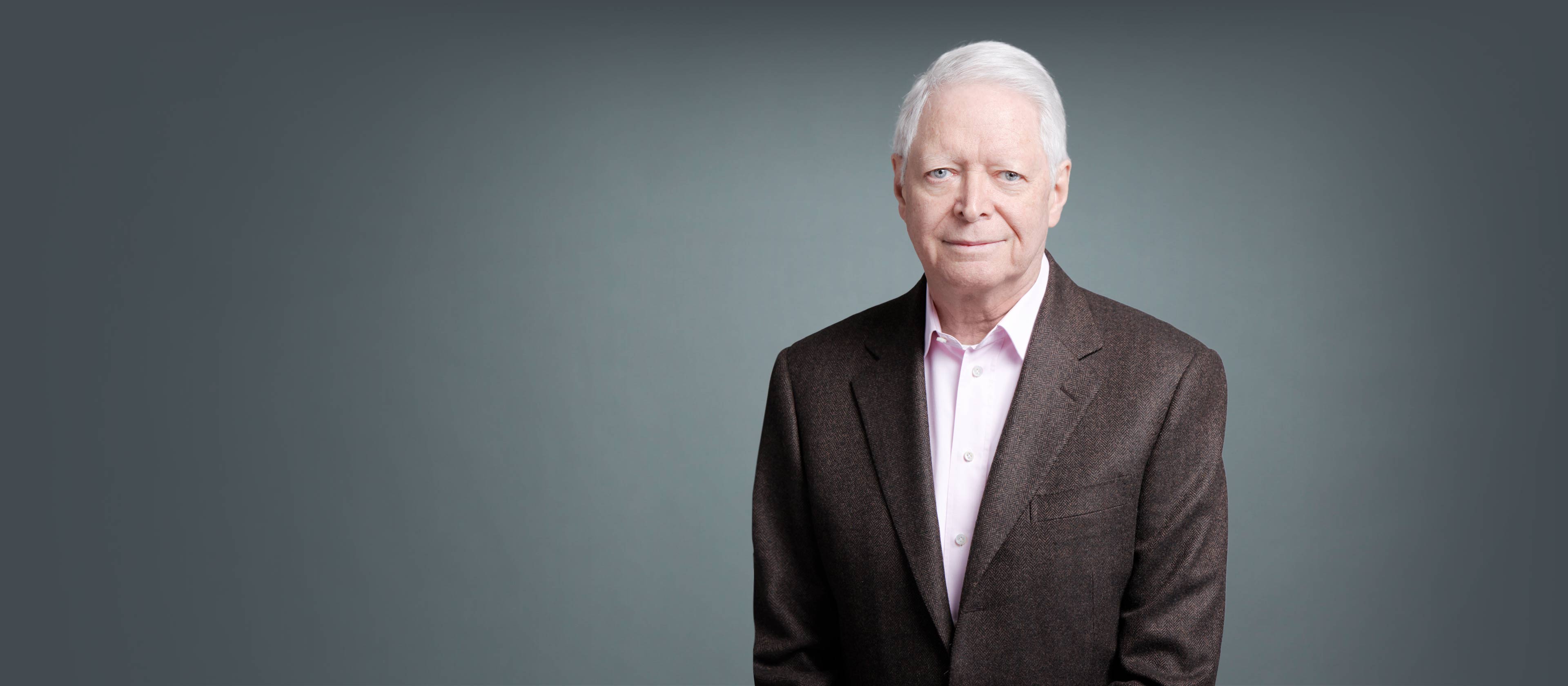
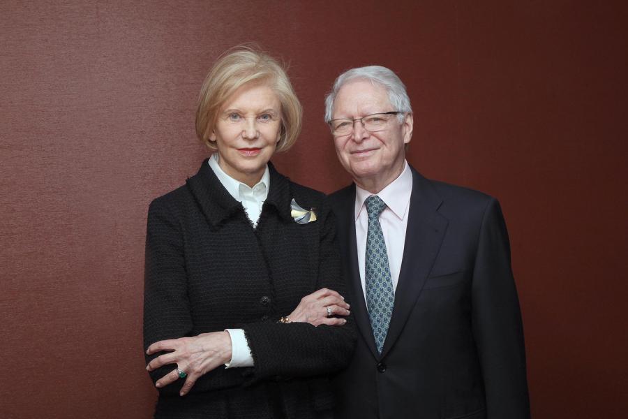
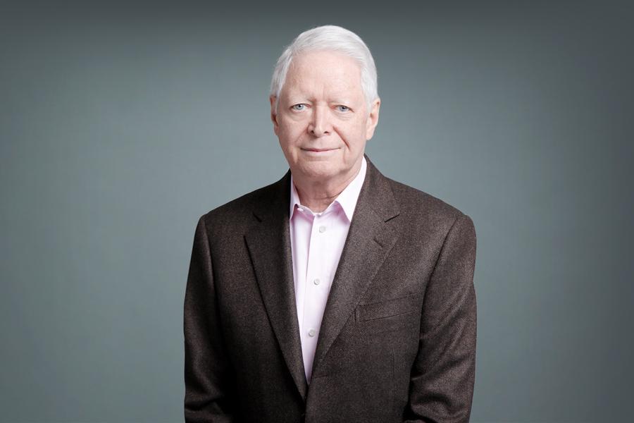
Research Professor, Department of Microbiology
Professor Emeritus of Microbiology, Department of Microbiology
Our research primarily concerns the mechanisms of cytokine actions, especially those of tumor necrosis factor (TNF) and the interferons (IFNs).
212-263-6756
212-263-9180
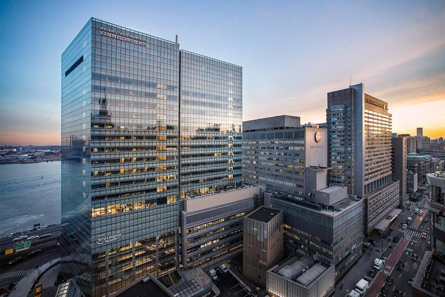
522 First Avenue
10, 1001
New York, NY 10016
Research Professor, Department of Microbiology at NYU Grossman School of Medicine
Professor Emeritus of Microbiology, Department of Microbiology at NYU Grossman School of Medicine
Fellowship, Institute of Virology, Czechoslovak Academy of Sciences, Bratislava, Virology
Institute of Virology, Czechoslovak Academy of Sciences, Bratislava, Virology
Journal of interferon & cytokine research. 2021 Apr; 41(4):137-138
Cell. 2020 Nov 12; 183(4):841-844
Proceedings of the National Academy of Sciences of the United States of America (PNAS). 2020 May 05; 117(18):9660-9664
Nature immunology. 2019 Jul; 20(7):775
Proceedings of the National Academy of Sciences of the United States of America (PNAS). 2019 Apr 09; 116(15):7157-7159
Proceedings of the National Academy of Sciences of the United States of America (PNAS). 2018 04 24; 115(17):4301-4304
Proceedings of the National Academy of Sciences of the United States of America (PNAS). 2017 02 21; 114(8):1748-1752