Main content
Adjunct Professor, Department of Radiology
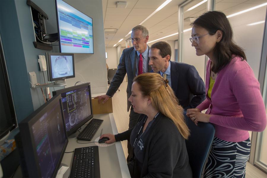
My research is primarily addressed at the development of new techniques for biomedical imaging. The broad aim of this work is to see what has previously been invisible in order to improve human health. I lead a multidisciplinary team that aims to develop a new paradigm of rapid continuous comprehensive imaging, taking advantage of complementary tools in image acquisition and reconstruction, including parallel imaging, compressed sensing, and artificial intelligence. This work extends to multiple imaging modalities, including MRI, PET and CT, and involves stakeholders in basic imaging science, clinical science, and industry, who work together in tightly coordinated method development and clinical evaluation.
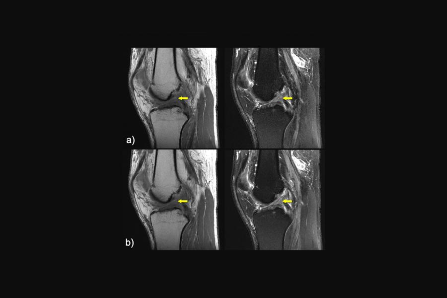
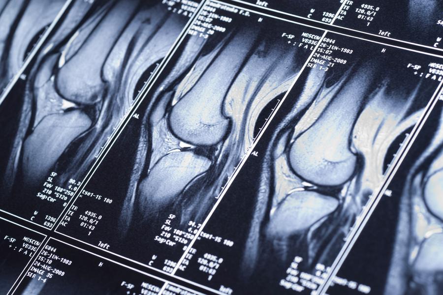
Early in my career, I played a leading role in the genesis of “parallel MRI”: the use of radiofrequency (RF) coil arrays to acquire MRI data in parallel rather than in a traditional sequential fashion, thereby enabling imaging at previously inaccessible speeds. Rapid MR imaging remains an area of strong interest for me, and my research group has contributed both to basic development and to multifaceted clinical implementations of rapid imaging techniques and technologies. This work combines the use of unconventional data sampling strategies with the development of novel algorithms for image reconstruction, incorporating recent advances in inverse problem regularization, deep learning, etc. For example, we are combining compressed sensing with parallel imaging and, more recently, machine learning in the pursuit of a few-minute comprehensive MR examination, which would enable rapid and robust diagnostic evaluations with rich information content and extremely simple workflow. We are also pursuing substantial reductions in CT radiation dose using methods translated from our research in rapid MRI. Another area of interest that derives originally from my work in parallel imaging is the design, optimization, and use of RF transmitters and detectors for high-performance MRI. As an outcome of this work, I have developed an ongoing interest in the interaction of electromagnetic fields with tissue, including perturbations resulting from tissue electrical properties, and potential new modalities for imaging of these electrical properties, which have previously eluded accurate noninvasive mapping. Both personally and in my capacity as Director of the Bernard and Irene Schwartz Center for Biomedical Imaging at NYU, I also have a strong interest in MR imaging and spectroscopy at high magnetic field strengths.
As Vice-Chair for Research in the Department of Radiology, I have overseen the development of a multifaceted translational imaging research program. This has culminated in the founding of our Center for Advanced Imaging Innovation and Research (CAI2R), which has operated since 2014 as an NIH Biomedical Technology Resource Center funded by the National Institute for Biomedical Imaging and Bioengineering. CAI2R develops novel imaging technologies for the improved management of cancer, musculoskeletal disease, and neurological disease, employing a unique new model for interdepartmental and academic-industrial collaboration to translate those technologies rapidly into clinical practice. In addition to ongoing work on rapid imaging and RF field interactions with tissue, one cornerstone of CAI2R research involves exploiting connections between imaging modalities such as MRI and PET (e.g. taking advantage of a combined MR-PET scanner operating in our Center), so as to advance the fundamental capabilities of each. In general, CAI2R aims at a new use of time in imaging, deploying leading-edge methods of rapid image acquisition and advanced image reconstruction to replace traditional complex and inefficient imaging protocols with simple, comprehensive, volumetric acquisitions that contain rich information about multiple complementary biophysical processes. Our overarching goal in CAI2R is to change the paradigms of data acquisition, image reconstruction, and day-to-day scanning in biomedical imaging, for the benefit of patients and the physicians who care for them.
212-263-4844
212-263-4845
660 First Avenue
Fourth Floor, Room 407
New York, NY 10016
Adjunct Professor, Department of Radiology at NYU Grossman School of Medicine
MD from Harvard Medical School
PhD from Massachusetts Institute of Technology
Beth Israel Deaconess Medical Center, Harvard Medical School, Boston, MA, Magnetic Resonance Imaging
Magnetic resonance in medicine. 2026 Mar; 95(3):1847-1857
Investigative radiology. 2026 Feb 01; 61(2):127-135
Nature communications. 2026 Jan 07;
Journal of magnetic resonance imaging. 2025 Dec 05;
Investigative radiology. 2025 Oct 01; 60(10):658-668
Journal of magnetic resonance imaging. 2025 Sep; 62(3):858-866
Human brain mapping. 2025 Jul; 46(10):e70232