Main content

Professor, Department of Neurosurgery
Professor, Department of Neuroscience
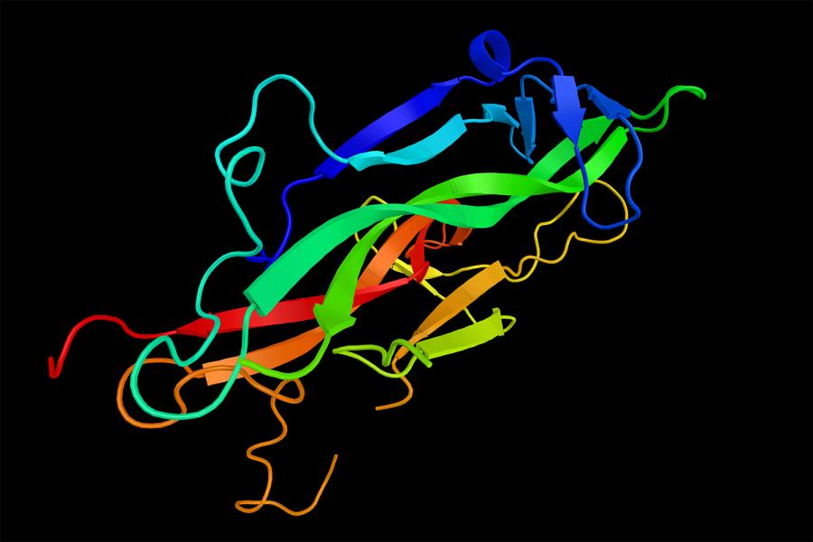
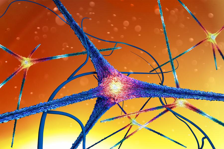
Dr. Rice's NIH-funded laboratory studies factors that regulate the release of dopamine, which is a key transmitter in motor and reward pathways of the brain. Current topics include modulation of dopamine release in the striatum by diet and by the metabolic hormones insulin and leptin, the influence of exercise on dopamine levels and release, and how a Parkinson's-related protein, alpha-synuclein, affects the physiology of dopamine neurons in the substantia nigra. Methods used include fast-scan cyclic voltammetry, optogenetics, patch-clamp recording of basal ganglia neurons, and immunohistochemistry. Dr. Rice is an investigator in the Neuroscience Institute and a member of the Marlene and Paolo Fresco Institute for Parkinson’s and Movement Disorders at NYU Langone, and she serves on the Scientific Advisory Board of the Parkinson’s Foundation.
212-263-5438
455 First Avenue
Eighth Floor, Suite 869
New York, NY 10016
Vice Chair, Research, Department of Neurosurgery
PhD from University of Kansas
Fellowship, New York University School of Medicine, Physiology and Biophysics
European journal of neuroscience. 2019 Mar; 49(6):794-804
ACS chemical neuroscience. 2017 02 15; 8(2):310-319
Nature communications. 2015 Oct 27; 6:8543
Basal ganglia. 2016 Aug; 6(3):123-148
NPJ Parkinson's disease. 2025 Dec 09; 11(1):345
Journal of neuroscience. 2025 Mar 24; 45(21):
Brain. 2024 Dec 03; 147(12):4017-4025
Analyst. 2024 Apr 15; 149(8):2351-2362