Contact Info
Address
140 Old Orangeburg Road
Orangeburg ,
NY
10962
845-398-5547
Education
— 12/91-12/92 Postdoctoral Fellow at the Pittsburgh Cancer Institute in Dr. Eli Gorelick's lab at Department of Pathology, PostDoctoral Training— Indian Institute of Science, Graduate Education
— 1/93-6/95 Postdoctoral Fellow in lab of late Prof. John P. Merlie, Department of Molecular Biology and Pharmacology, Washington University School of Medicine, PostDoctoral Training
— 7/95-7/99 Postdoctoral fellow in the laboratory of Dr. Don W. Cleveland, Department of Cell Biology, UCSD School of Medicine, PostDoctoral Training
— 8/86-11/91 Graduate student at the Department of Biochemistry in Prof. G. Padmanaban's lab, PostDoctoral Training
Research Summary
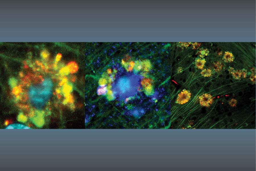
The major focus of my lab is to understand molecular cues that control the radia l growth of axons and death of motor neurons. Radial growth of axons is studied in mice by ES cell mediated homologous recombination. Motor neurons disease is s tudied in mouse models of amyotrophic lateral sclerosis (ALS) in transgenic mice for neurofilaments and superoxide dismutase 1 (SOD1). During development, axons undergo two major changes. First, the long, thin axons make growth cones and establish stable synapses. Second, axonal volume increase s 100 fold, and large myelinated axons accumulate large numbers of neurofilament s (NFs). NFs are 10 nm filaments composed of NF-H (200 kd), NF-M (150 kd) and N F-L (68 kd) subunits. Genetic analysis has shown a direct correlation between t he number of filaments and axonal volume. Our transgenic mouse models have indi cated that subunit ratios play a critical role in controlling radial growth of a xons. Recent gene deletion analysis on individual subunits indicate that NF-L i s important for filament formation; NF-M is important for filament assembly and control of filament number. In order to identify the domains of NF subunits tha t are responsible for controlling the radial growth of axons, a systematic domai n deletion approach is being used. We have successfully produced mice for carbox yl terminal deletions of NF-M and NF-H, and analyses of these mice are in progre ss. Approximately 2% of human ALS patients have mutations in the gene encoding for S OD1. A hallmark of ALS is neurofilament accumulation in cell bodies and proxima l axons. Over 50 mutations in SOD1 have been identified in human ALS patients. A large number of mouse models exist for ALS. The role of each NF subunit in d isease onset and progression of motor neuron death is unknown. In order to addr ess this question, NF-H deleted and over-expressing mice are bred with SOD1 over -expressing mice and efforts are underway to characterize these mice. Results will indicate the role of NF-H in motor neuron-mediated cell death.All data from NYU Health Sciences Library Faculty Bibliography — -
Contact:
http://hsl.med.nyu.edu/faculty-bibliography-search#about
The Myosin Va Head Domain Binds to the Neurofilament-L Rod and Modulates Endoplasmic Reticulum (ER) Content and Distribution within Axons
Rao, Mala V; Mohan, Panaiyur S; Kumar, Asok; Yuan, Aidong; Montagna, Lee; Campbell, Jabbar; Espreafico, Enilza M; Julien, Jean P; Nixon, Ralph A
2011 ;6(2):e17087-e17087, PLoS ONE
The neurofilament light subunit (NF-L) binds to myosin Va (Myo Va) in neurons but the sites of interaction and functional significance are not clear. We show by deletion analysis that motor domain of Myo Va binds to the NF-L rod domain that forms the NF backbone. Loss of NF-L and Myo Va binding from axons significantly reduces the axonal content of ER, and redistributes ER to the periphery of axon. Our data are consistent with a novel function for NFs as a scaffold in axons for maintaining the content and proper distribution of vesicular organelles, mediated in part by Myo Va. Based on observations that the Myo Va motor domain binds to intermediate filament (IF) proteins of several classes, Myo Va interactions with IFs may serve similar roles in organizing organelle topography in different cell types
—
id: 126479,
year: 2011,
vol: 6,
page: e17087,
stat: Journal Article,
The contributions of myelin and axonal caliber to transverse relaxation time in shiverer and neurofilament-deficient mouse models
Dyakin, Victor V; Chen, Yuanxin; Branch, Craig A; Yuan, Aidong; Rao, Mala; Kumar, Asok; Peterhoff, Corrinne M; Nixon, Ralph A
2010 Jul 1;51(3):1098-1105, Neuroimage
White matter disorders can involve injury to myelin or axons but the respective contribution of each to clinical course is difficult to evaluate non-invasively. Here, to develop a paradigm for further investigations of axonal pathology by MRI, we compared two genetic mouse models exhibiting relatively selective axonal or myelin deficits using quantitative MRI relaxography of the transverse relaxation times (T2) in vivo and ultrastructural morphometry. In HM-DKO mice, which lack genes encoding the heavy (NF-H) and medium (NF-M) subunits of neurofilaments, neurofilament content of large myelinated axons of the central nervous system (CNS) is markedly reduced in the absence of changes in myelin thickness and volume. In shiverer mutant mice, which lack functional myelin basic protein, CNS myelin sheath formation is markedly reduced but neurofilament content is normal. We observed increases in T2 in nearly all white matter in shiverer mice compared to their wild type, while more subtle increases in T2 were observed in HM-DKO in the corpus callosum. White matter T2 was generally greater in shiverer mice than HM-DKO mice. Ultrastructural morphometry of the corpus callosum, which exhibited the greatest T2 differences, confirmed that total cross-sectional area occupied by axons was similar in the two mouse models and that the major ultrastructural differences, determined by morphometry, were an absence of myelin and larger unmyelinated axons in shiverer mice and absence of neurofilaments in HM-DKO mice. Our findings indicate that T2 is strongly influenced by myelination state and axonal volume, while neurofilament structure within the intra-axonal compartment has a lesser effect upon single compartment T2 estimates
—
id: 126489,
year: 2010,
vol: 51,
page: 1098,
stat: Journal Article,
Phosphorylation of highly conserved neurofilament medium KSP repeats is not required for myelin-dependent radial axonal growth
Garcia, Michael L; Rao, Mala V; Fujimoto, Jiro; Garcia, Virginia B; Shah, Sameer B; Crum, John; Gotow, Takahiro; Uchiyama, Yasuo; Ellisman, Mark; Calcutt, Nigel A; Cleveland, Don W
2009 Feb 4;29(5):1277-1284, Journal of neuroscience
Neurofilament medium (NF-M) is essential for the acquisition of normal axonal caliber in response to a myelin-dependent 'outside-in' trigger for radial axonal growth. Removal of the tail domain and lysine-serine-proline (KSP) repeats of NF-M, but not neurofilament heavy, produced axons with impaired radial growth and reduced conduction velocities. These earlier findings supported myelin-dependent phosphorylation of NF-M KSP repeats as an essential component of axonal growth. As a direct test of whether phosphorylation of NF-M KSP repeats is the target for the myelin-derived signal, gene replacement has now been used to produce mice in which all serines of NF-M's KSP repeats have been replaced with phosphorylation-incompetent alanines. This substitution did not alter accumulation of the neurofilaments or their subunits. Axonal caliber and motor neuron conduction velocity of mice expressing KSP phospho-incompetent NF-M were also indistinguishable from wild-type mice. Thus, phosphorylation of NF-M KSP repeats is not an essential component for the acquisition of normal axonal caliber mediated by myelin-dependent outside-in signaling
—
id: 94361,
year: 2009,
vol: 29,
page: 1277,
stat: Journal Article,
PHOSPHORYLATION OF NF-M KSP REPEATS IS NOT REQUIRED FOR RADIAL AXONAL GROWTH
Garcia, ML; Rao, MV; Garcia, VB; Ellisman, M; Calcutt, NA; Cleveland, DW
2009 JUL ;14(9):54-55, Journal of the peripheral nervous system
—
id: 102289,
year: 2009,
vol: 14,
page: 54,
stat: Journal Article,
Neurofilaments form a highly stable stationary cytoskeleton after reaching a critical level in axons
Yuan, Aidong; Sasaki, Takahiro; Rao, Mala V; Kumar, Asok; Kanumuri, Vivek; Dunlop, David S; Liem, Ronald K; Nixon, Ralph A
2009 Sep 9;29(36):11316-11329, Journal of neuroscience
The ultrastructural view of the axonal cytoskeleton as an extensively cross-linked network of neurofilaments (NFs) and other cytoskeletal polymers contrasts with the dynamic view suggested by axonal transport studies on cytoskeletal elements. Here we reconcile these perspectives by showing that neurons form a large NF network along axons which is unequivocally stationary, metabolically stable, and maintained by NFs and nonfilamentous subunit assemblies undergoing slow transport by intermittent rapid movements and pauses. In mouse primary cortical neurons transfected with EGFP-NFL, formation of this stationary NF network requires a critical level of NFs, which explains its absence in NF-poor developing neurons studied previously. Most NFs at proximal axon regions were in a stationary structure coexisting with a smaller pool of moving EGFP-NFL assemblies that were mainly nonfilamentous. Distally along the same axon, EGFP-labeled NFL was much less abundant, and we detected only short filaments moving bidirectionally by slow transport (rapid movements and pauses) as previously described. In living mice, >25% of radiolabeled newly synthesized NFs remained in optic axons after slowly transported NFs had exited. Retained NF remained fixed over several months in a nonuniform distribution and exhibited exceptionally slow turnover (t(1/2) >2.5 months), implying that, at steady state, >90% of NFs in mature optic axons comprise the stationary cytoskeleton and <10% are undergoing slow transport. These findings reconcile in vitro and in vivo axonal transport observations, showing that slowly transported NFs or subunit oligomers are precursors to a highly stable stationary cytoskeletal network that supports mature axons
—
id: 126492,
year: 2009,
vol: 29,
page: 11316,
stat: Journal Article,
Marked calpastatin (CAST) depletion in Alzheimer's disease accelerates cytoskeleton disruption and neurodegeneration: neuroprotection by CAST overexpression
Rao, Mala V; Mohan, Panaiyur S; Peterhoff, Corrinne M; Yang, Dun-Sheng; Schmidt, Stephen D; Stavrides, Philip H; Campbell, Jabbar; Chen, Yuanxin; Jiang, Ying; Paskevich, Peter A; Cataldo, Anne M; Haroutunian, Vahram; Nixon, Ralph A
2008 Nov 19;28(47):12241-12254, Journal of neuroscience
Increased activity of calpains is implicated in synaptic dysfunction and neurodegeneration in Alzheimer's disease (AD). The molecular mechanisms responsible for increased calpain activity in AD are not known. Here, we demonstrate that disease progression is propelled by a marked depletion of the endogenous calpain inhibitor, calpastatin (CAST), from AD neurons, which is mediated by caspase-1, caspase-3, and calpains. Initial CAST depletion focally along dendrites coincides topographically with calpain II and ERK 1/2 activation, tau cleavage by caspase-3, and tau and neurofilament hyperphosphorylation. These same changes, together with cytoskeletal proteolysis and neuronal cell death, accompany CAST depletion after intrahippocampal kainic acid administration to mice, and are substantially reduced in mice overexpressing human CAST. Moreover, CAST reduction by shRNA in neuronal cells causes calpain-mediated death at levels of calcium-induced injury that are sublethal to cells normally expressing CAST. Our results strongly support a novel hypothesis that CAST depletion by multiple abnormally activated proteases accelerates calpain dysregulation in AD leading to cytoskeleton disruption and neurodegeneration. CAST mimetics may, therefore, be neuroprotective in AD
—
id: 94362,
year: 2008,
vol: 28,
page: 12241,
stat: Journal Article,
Deleting the phosphorylated tail domain of the neurofilament heavy subunit does not alter neurofilament transport rate in vivo
Yuan, Aidong; Nixon, Ralph A; Rao, Mala V
2006 Jan 30;393(2-3):264-268, Neuroscience letters
Phosphorylation of the carboxyl tail domains of the neurofilament heavy (NF-H) and middle molecular weight (NF-M) subunits has been proposed to regulate the axonal transport of neurofilaments. To test this hypothesis, we recently constructed mice lacking the extensively phosphorylated NF-H tail domain (NF-HtailDelta) and showed that the transport rate of neurofilaments in optic axons is unaltered in the absence of this domain [M.V. Rao, M.L. Garcia, Y. Miyazaki, T. Gotow, A. Yuan, S. Mattina, C.M. Ward, N.A. Calcutt, Y. Uchiyama, R.A. Nixon, D.W. Cleveland, Gene replacement in mice reveals that the heavily phosphorylated tail of neurofilament heavy subunit does not affect axonal caliber or the transit of cargoes in slow axonal transport, J. Cell Biol. 158 (2002) 681-693]. However, Shea et al. proposed that deletion of NF-H carboxyl-terminal region accelerates the transport of a subpopulation of neurofilaments based on minor differences between tail-deleted and control mice in our axonal transport analysis. Here, we present additional evidence that neurofilament transport rate is unchanged after deleting the phosphorylated NF-H tail domain, establishing unequivocally that the NF-H tail domain alone does not regulate the rate of neurofilament transport in optic axons in vivo. Possible roles for tail domains as cross-bridges between a neurofilament and its neighbors or other cytoskeletal elements is discussed
—
id: 60252,
year: 2006,
vol: 393,
page: 264,
stat: Journal Article,
Alpha-internexin is structurally and functionally associated with the neurofilament triplet proteins in the mature CNS
Yuan, Aidong; Rao, Mala V; Sasaki, Takahiro; Chen, Yuanxin; Kumar, Asok; Liem, Ronald K H; Eyer, Joel; Peterson, Alan C; Julien, Jean-Pierre; Nixon, Ralph A
2006 Sep 27;26(39):10006-10019, Journal of neuroscience
Alpha-internexin, a neuronal intermediate filament protein implicated in neurodegenerative disease, coexists with the neurofilament (NF) triplet proteins (NF-L, NF-M, and NF-H) but has an unknown function. The earlier peak expression of alpha-internexin than the triplet during brain development and its ability to form homopolymers, unlike the triplet, which are obligate heteropolymers, have supported a widely held view that alpha-internexin and neurofilament triplet form separate filament systems. Here, we demonstrate, however, that despite a postnatal decline in expression, alpha-internexin is as abundant as the triplet in the adult CNS and exists in a relatively fixed stoichiometry with these subunits. Alpha-internexin exhibits transport and turnover rates identical to those of triplet proteins in optic axons and colocalizes with NF-M on single neurofilaments by immunogold electron microscopy. Alpha-internexin also coassembles with all three neurofilament proteins into a single network of filaments in quadruple-transfected SW13vim(-) cells. Genetically deleting NF-M alone or together with NF-H in mice dramatically reduces alpha-internexin transport and content in axons throughout the CNS. Moreover, deleting alpha-internexin potentiates the effects of NF-M deletion on NF-H and NF-L transport. Finally, overexpressing a NF-H-LacZ fusion protein in mice induces alpha-internexin and neurofilament triplet to aggregate in neuronal perikarya and greatly reduces their transport and content selectively in axons. Our data show that alpha-internexin and the neurofilament proteins are functionally interdependent. The results strongly support the view that alpha-internexin is a fourth subunit of neurofilaments in the adult CNS, providing a basis for its close relationship with neurofilaments in CNS diseases associated with neurofilament accumulation
—
id: 94108,
year: 2006,
vol: 26,
page: 10006,
stat: Journal Article,
The neurofilament middle molecular mass subunit carboxyl-terminal tail domains is essential for the radial growth and cytoskeletal architecture of axons but not for regulating neurofilament transport rate
Rao, Mala V; Campbell, Jabbar; Yuan, Aidong; Kumar, Asok; Gotow, Takahiro; Uchiyama, Yasuo; Nixon, Ralph A
2003 Dec 8;163(5):1021-1031, Journal of cell biology
The phosphorylated carboxyl-terminal 'tail' domains of the neurofilament (NF) subunits, NF heavy (NF-H) and NF medium (NF-M) subunits, have been proposed to regulate axon radial growth, neurofilament spacing, and neurofilament transport rate, but direct in vivo evidence is lacking. Because deletion of the tail domain of NF-H did not alter these axonal properties (Rao, M.V., M.L. Garcia, Y. Miyazaki, T. Gotow, A. Yuan, S. Mattina, C.M. Ward, N.S. Calcutt, Y. Uchiyama, R.A. Nixon, and D.W. Cleveland. 2002. J. Cell Biol. 158:681-693), we investigated possible functions of the NF-M tail domain by constructing NF-M tail-deleted (NF-MtailDelta) mutant mice using an embryonic stem cell-mediated 'gene knockin' approach that preserves normal ratios of the three neurofilament subunits. Mutant NF-MtailDelta mice exhibited severely inhibited radial growth of both motor and sensory axons. Caliber reduction was accompanied by reduced spacing between neurofilaments and loss of long cross-bridges with no change in neurofilament protein content. These observations define distinctive functions of the NF-M tail in regulating axon caliber by modulating the organization of the neurofilament network within axons. Surprisingly, the average rate of axonal transport of neurofilaments was unaltered despite these substantial effects on axon morphology. These results demonstrate that NF-M tail-mediated interactions of neurofilaments, independent of NF transport rate, are critical determinants of the size and cytoskeletal architecture of axons, and are mediated, in part, by the highly phosphorylated tail domain of NF-M
—
id: 40064,
year: 2003,
vol: 163,
page: 1021,
stat: Journal Article,
Defective neurofilament transport in mouse models of amyotrophic lateral sclerosis: a review
Rao, Mala V; Nixon, Ralph A
2003 Jul;28(7):1041-1047, Neurochemical research
Neurofilament proteins synthesized in the cell body of neurons are assembled and transported into axons, where they influence axon radial growth, axonal transport, and nerve conduction velocities. In diseased states, neurofilaments accumulate in cell bodies and proximal axons of affected neurons, and these lesions are characteristic of many neurodegenerative diseases, including amyotrophic lateral sclerosis (ALS), Alzheimer's disease (AD), spinal muscular atrophy (SMA), Charcot-Marie-Tooth disease type 2 (CMT2), and hereditary sensory motor neuropathy. Although the molecular mechanisms that contribute to these accumulations are not yet identified, transgenic mouse models are beginning to provide insight into the role of neurofilament transport in disease-related dysfunction of neurons. This review addresses axonal transport in mouse models of ALS and the special significance of neurofilament transport in this disease
—
id: 60267,
year: 2003,
vol: 28,
page: 1041,
stat: Journal Article,
Neurofilament transport in vivo minimally requires hetero-oligomer formation
Yuan, Aidong; Rao, Mala V; Kumar, Asok; Julien, Jean-Pierre; Nixon, Ralph A
2003 Oct 15;23(28):9452-9458, Journal of neuroscience
Neurofilament assembly requires at minimum the polymerization of neurofilament light chain (NF-L) with either neurofilament medium chain (NF-M) or neurofilament heavy chain (NF-H) subunits, but requirements for their axonal transport have long been controversial. Using a gene deletion approach, we generated mice containing only NF-L or NF-M. In vivo pulse radiolabeling analyses in retinal ganglion cell neurons revealed that NF-L alone is incapable of efficient transport, whereas nearly one-half of the normal level of NF-M is transported along optic axons in the absence of the other triplet subunits. Under these conditions, however, NF-M transport is completely abolished by deleting alpha-internexin. Our results strongly suggest that efficient neurofilament protein transport in vivo minimally requires hetero-oligomer formation. They also show that NF-M can partner with intermediate filament proteins other than the NF-H and NF-L subunits in neurons to support slow transport and possibly other functions of neuronal intermediate filaments
—
id: 39029,
year: 2003,
vol: 23,
page: 9452,
stat: Journal Article,
Myosin Va binding to neurofilaments is essential for correct myosin Va distribution and transport and neurofilament density
Rao, Mala V; Engle, Linda J; Mohan, Panaiyur S; Yuan, Aidong; Qiu, Dike; Cataldo, Anne; Hassinger, Linda; Jacobsen, Stephen; Lee, Virginia M-Y; Andreadis, Athena; Julien, Jean-Pierre; Bridgman, Paul C; Nixon, Ralph A
2002 Oct 28;159(2):279-290, Journal of cell biology
The identification of molecular motors that modulate the neuronal cytoskeleton has been elusive. Here, we show that a molecular motor protein, myosin Va, is present in high proportions in the cytoskeleton of mouse CNS and peripheral nerves. Immunoelectron microscopy, coimmunoprecipitation, and blot overlay analyses demonstrate that myosin Va in axons associates with neurofilaments, and that the NF-L subunit is its major ligand. A physiological association is indicated by observations that the level of myosin Va is reduced in axons of NF-L-null mice lacking neurofilaments and increased in mice overexpressing NF-L, but unchanged in NF-H-null mice. In vivo pulse-labeled myosin Va advances along axons at slow transport rates overlapping with those of neurofilament proteins and actin, both of which coimmunoprecipitate with myosin Va. Eliminating neurofilaments from mice selectively accelerates myosin Va translocation and redistributes myosin Va to the actin-rich subaxolemma and membranous organelles. Finally, peripheral axons of dilute-lethal mice, lacking functional myosin Va, display selectively increased neurofilament number and levels of neurofilament proteins without altering axon caliber. These results identify myosin Va as a neurofilament-associated protein, and show that this association is essential to establish the normal distribution, axonal transport, and content of myosin Va, and the proper numbers of neurofilaments in axons
—
id: 32536,
year: 2002,
vol: 159,
page: 279,
stat: Journal Article,
Gene replacement in mice reveals that the heavily phosphorylated tail of neurofilament heavy subunit does not affect axonal caliber or the transit of cargoes in slow axonal transport
Rao, Mala V; Garcia, Michael L; Miyazaki, Yukio; Gotow, Takahiro; Yuan, Aidong; Mattina, Salvatore; Ward, Chris M; Calcutt, Nigel A; Uchiyama, Yasuo; Nixon, Ralph A; Cleveland, Don W
2002 Aug 19;158(4):681-693, Journal of cell biology
The COOH-terminal tail of mammalian neurofilament heavy subunit (NF-H), the largest neurofilament subunit, contains 44-51 lysine-serine-proline repeats that are nearly stoichiometrically phosphorylated after assembly into neurofilaments in axons. Phosphorylation of these repeats has been implicated in promotion of radial growth of axons, control of nearest neighbor distances between neurofilaments or from neurofilaments to other structural components in axons, and as a determinant of slow axonal transport. These roles have now been tested through analysis of mice in which the NF-H gene was replaced by one deleted in the NF-H tail. Loss of the NF-H tail and all of its phosphorylation sites does not affect the number of neurofilaments, alter the ratios of the three neurofilament subunits, or affect the number of microtubules in axons. Additionally, it does not reduce interfilament spacing of most neurofilaments, the speed of action potential propagation, or mature cross-sectional areas of large motor or sensory axons, although its absence slows the speed of acquisition of normal diameters. Most surprisingly, at least in optic nerve axons, loss of the NF-H tail does not affect the rate of transport of neurofilament subunits
—
id: 32535,
year: 2002,
vol: 158,
page: 681,
stat: Journal Article,
Progress in the modeling of neurodegenerative diseases in transgenic mice
Duff K; Rao MV
2001 Aug;14(4):441-447, Current opinion in neurology
Transgenic mouse models exist for the major neurodegenerative diseases, including Alzheimer's disease, tauopathy and amyotrophic lateral sclerosis. Although many of the mice do not completely replicate the human disease they are intended to model, they have provided insight into the mechanisms that underlie disease etiology. In the case of the Alzheimer's disease and amyotrophic lateral sclerosis models, the mice have also provided a therapeutic testing ground for the testing of agents that have been shown to have considerable clinical promise
—
id: 24731,
year: 2001,
vol: 14,
page: 441,
stat: Journal Article,
Neurofilaments
Nixon RA; Rao MV
Encyclopedia of molecular medicine Chichester : Wiley, 2001,
—
id: 2724,
year: 2001,
vol: ,
page: 1589,
stat: Chapter,
Neurofilament-dependent radial growth of motor axons and axonal organization of neurofilaments does not require the neurofilament heavy subunit (NF-H) or its phosphorylation
Rao MV; Houseweart MK; Williamson TL; Crawford TO; Folmer J; Cleveland DW
1998 Oct 5;143(1):171-181, Journal of cell biology
Neurofilaments are essential for establishment and maintenance of axonal diameter of large myelinated axons, a property that determines the velocity of electrical signal conduction. One prominent model for how neurofilaments specify axonal growth is that the 660-amino acid, heavily phosphorylated tail domain of neurofilament heavy subunit (NF-H) is responsible for neurofilament-dependent structuring of axoplasm through intra-axonal crossbridging between adjacent neurofilaments or to other axonal structures. To test such a role, homologous recombination was used to generate NF-H-null mice. In peripheral motor and sensory axons, absence of NF-H does not significantly affect the number of neurofilaments or axonal elongation or targeting, but it does affect the efficiency of survival of motor and sensory axons. Loss of NF-H caused only a slight reduction in nearest neighbor spacing of neurofilaments and did not affect neurofilament distribution in either large- or small-diameter motor axons. Since postnatal growth of motor axon caliber continues largely unabated in the absence of NF-H, neither interactions mediated by NF-H nor the extensive phosphorylation of it within myelinated axonal segments are essential features of this growth
—
id: 24732,
year: 1998,
vol: 143,
page: 171,
stat: Journal Article,
Distinct regulatory elements control muscle-specific, fiber-type-selective, and axially graded expression of a myosin light-chain gene in transgenic mice
Rao MV; Donoghue MJ; Merlie JP; Sanes JR
1996 Jul;16(7):3909-3922, Molecular & cellular biology
The fast alkali myosin light chain 1f/3f (MLC1f/3f) gene is developmentally regulated, muscle specific, and preferentially expressed in fast-twitch fibers. A transgene containing an MLC1f promoter plus a downstream enhancer replicates this pattern of expression in transgenic mice. Unexpectedly, this transgene is also expressed in a striking (approximately 100-fold) rostrocaudal gradient in axial muscles (reviewed by J. R. Sanes, M. J. Donoghue, M. C. Wallace, and J. P. Merlie, Cold Spring Harbor Symp. Quant. Biol. 57:451-460, 1992). Here, we analyzed the expression of mutated transgenes to map sites necessary for muscle-specific, fiber-type-selective, and axially graded expression. We show that two E boxes (myogenic factor binding sites), a homeodomain (hox) protein binding site, and an MEF2 site, which are clustered in an approximately 170-bp core enhancer, are all necessary for maximal transgene activity in muscle but not for fiber-type- or position-dependent expression. A distinct region within the core enhancer promotes selective expression of the transgene in fast-twitch muscles. Sequences that flank the core enhancer are also necessary for high-level activity in transgenic mice but have little influence on activity in transfected cells, suggesting the presence of regions resembling matrix attachment sites. Truncations of the MLC1f promoter affected position-dependent expression of the transgene, revealing distinct regions that repress transgene activity in neck muscles and promote differential expression among intercostal muscles. Thus, the whole-body gradient of expression displayed by the complete transgene may reflect the integrated activities of discrete elements that regulate expression in subsets of muscles. Finally, we show that transgene activity is not significantly affected by deletion or overexpression of the myoD gene, suggesting that intermuscular differences in myogenic factor levels do not affect patterns of transgene expression. Together, our results provide evidence for at least nine distinct sites that exert major effects on the levels and patterns of MLC1f expression in adult muscles
—
id: 24766,
year: 1996,
vol: 16,
page: 3909,
stat: Journal Article,
Molecular docking programs successfully predict the binding of a beta-lactamase inhibitory protein to TEM-1 beta-lactamase
Strynadka, NCJ; Eisenstein, M; KatchalskiKatzir, E; Shoichet, BK; Kuntz, ID; Abagyan, R; Totrov, M; Janin, J; Cherfils, J; Zimmerman, F; Olson, A; Duncan, B; Rao, M; Jackson, R; Sternberg, M; James, MNG
1996 MAR ;3(3):233-239, Nature structural biology
Crystallization of the 1:1 molecular complex between the beta-lactamase TEM-1 and the beta-lactamase inhibitory protein BLIP has provided an opportunity to put a stringent test on current protein-docking algorithms, Prior to the successful determination of the structure of the complex, nine laboratory groups were given the refined atomic coordinates of each of the native molecules, Other than the fact that BLIP is an effective inhibitor of a number of beta-lactamase enzymes (KI for TEM-1 similar to 100 pM) no other biochemical or structural data were available to assist the practitioners in their molecular docking, In addition, it was not known whether the molecules underwent conformational changes upon association or whether the inhibition was competitive or non-competitive. All six of the groups that accepted the challenge correctly predicted the general mode of association of BLIP and TEM-1
—
id: 53051,
year: 1996,
vol: 3,
page: 233,
stat: Journal Article,
Characterization of a negative cis-acting DNA element regulating the transcription of CYP2B1/B2 gene in rat liver
Ram N; Rao MV; Prabhu L; Nirodi CS; Sultana S; Vatsala PG; Padmanaban G
1995 Feb 20;317(1):39-45, Archives of biochemistry & biophysics. ABB
The region -160 to -127 nt of the upstream of CYP2B1/B2 gene has been found to function as a negative cis-acting element on the basis of DNase-I footprint and gel mobility shift assays as well as cell-free transcriptional assays using Bal-31 mutants. A reciprocal relationship in the interaction of the negative and the recently characterized positive elements with their respective protein factors has been found under repressed and induced conditions of the gene. The negative element also harbors the core glucocorticoid responsive sequence, TGTCCT. It is concluded that the negative element mediates the repressed state of the gene under the uninduced condition and also mediates the repressive effect of dexamethasone, when given along with the inducer phenobarbitone in rats. Dexamethasone is able to antagonize the effects of phenobarbitone at as low a concentration as 100 micrograms/kg body wt in these animals
—
id: 24733,
year: 1995,
vol: 317,
page: 39,
stat: Journal Article,
Lectin-induced apoptosis of tumour cells
Kim M; Rao MV; Tweardy DJ; Prakash M; Galili U; Gorelik E
1993 Oct;3(5):447-453, Glycobiology
The mechanisms of cytotoxic activity of Griffonia simplicifolia 1-B4 (GS1B4) and wheat germ agglutinin (WGA) lectins against various murine tumour cell lines were studied. Tumour cells that lack lectin-binding carbohydrates were resistant to lysis by these lectins. However, YAC-1 cells that expressed GS1B4 lectin-binding sites showed low sensitivity to lysis. To further analyse the relative importance of cell surface carbohydrates in lectin cytotoxicity, BL6-8 melanoma cells, which do not express the alpha 1,3 galactosyltransferase (alpha 1,3GT) gene and cell surface alpha-galactosyl epitopes reacting with GS1B4 lectin, were transfected with cDNA encoding alpha 1,3GT. After transfection, BL6-8 cells expressed high levels of GS1B4-binding alpha-galactosyl epitopes, but remained resistant to lysis by GS1B4 lectin, suggesting that the presence of lectin-binding epitopes, while essential, is not sufficient for tumour cell lysis and probably some intracellular mechanisms are involved in the regulation of lectin-mediated cytotoxicity. We found that the GS1B4 and WGA lectins induced apoptosis with DNA fragmentation of sensitive, but not resistant, tumour cell lines. DNA fragmentation, as well as tumour cell lysis, was blocked in the presence of the specific inhibitory sugar. To determine whether binding of the lectin to cell surface carbohydrates is sufficient to trigger tumour cell lysis, lectin-sensitive CL8-1 melanoma cells were incubated with GS1B4 lectin immobilized on agarose beads. Although these tumour cells bind to the immobilized lectin, it failed to trigger tumour cell death, suggesting that only soluble lectin is capable of tumour cell lysis and lectin internalization is probably required for their lysis.(ABSTRACT TRUNCATED AT 250 WORDS)
—
id: 24734,
year: 1993,
vol: 3,
page: 447,
stat: Journal Article,
Identification and functional characterization of a cis-acting positive DNA element regulating CYP 2B1/B2 gene transcription in rat liver
Upadhya P; Rao MV; Venkateswar V; Rangarajan PN; Padmanaban G
1992 Feb 11;20(3):557-562, Nucleic acids research
A positive cis-acting DNA element in the near 5'-upstream region of the CYP2B1/B2 genes in rat liver was found to play an important role in the transcription of these genes. An oligonucleotide covering -69 to -98 nt mimicked the gel mobility shift pattern given by the fragment -179 to +29 nt, which was earlier found adequate to confer the regulatory features of this gene. Two major complexes were seen, of which the slower and faster moving complexes became intense under uninduced and Phenobarbitone-induced conditions respectively. Minigene cloned DNA plasmid covering -179 to +181 nt in pUC 19 and Bal 31 mutants derived from this parent were transcribed in whole nuclei and cell free transcription extracts and mutants containing only upto -75 nt of the upstream were poorly transcribed. Transcription extracts from phenobarbitone-injected rat liver nuclei were significantly more active than extracts from uninduced rats in transcribing the minigene constructs. Addition of the oligonucleotide (-69 to -98nt) specifically inhibited the transcription of the minigene construct (-179 to +181 nt) in the cell free transcription system. It is therefore, concluded that the region -69 to -98 nt acts as a positive cis-acting element in the transcription of the CYP2B1/B2 genes and in mediating the inductive effects of phenobarbitone
—
id: 24735,
year: 1992,
vol: 20,
page: 557,
stat: Journal Article,
Transcriptional regulation of Cytochrome P-450b/e gene expression in rat liver
Padmanaban G; Rangarajan PN; Venkateswar V; Rao MV; Upadya P
Biological oxidation systems San Diego : Academic Press, 1990,
—
id: 2659,
year: 1990,
vol: 1,
page: 39,
stat: Chapter,
Dexamethasone negatively regulates phenobarbitone-activated transcription but synergistically enhances cytoplasmic levels of cytochrome P-450b/e messenger RNA
Rao MV; Rangarajan PN; Padmanaban G
1990 Apr 5;265(10):5617-5622, Journal of biological chemistry
Dexamethasone has a potentiating effect on phenobarbitone mediated induction of cytochrome P-450b + e mRNAs in adult rat liver. However, the glucocorticoid inhibits phenobarbitone-activated transcription of cytochrome P-450b + e mRNAs by 60-70%. This inhibitory effect is evident in run-off transcription of the endogenous genes as well as in the transcription of an added cloned gene fragment. Dexamethasone inhibits the phenobarbitone-mediated increase in the binding of a transcription factor(s) to the upstream region of the gene as evidenced by gel retardation and Southwestern blot analysis. The glucocorticoid does not stabilize the phenobarbitone-induced polyribosomal cytochrome P-450b + e mRNAs but appears to stabilize the nuclear transcripts. It is proposed that a negative element may mediate the action of dexamethasone at the level of nuclear transcription and stabilization of the nuclear transcript may account for the potentiating effect of the glucocorticoid on phenobarbitone-mediated increase in cytochrome P-450b + e mRNAs in the cytoplasm of the adult rat liver. However, the cytochrome P-450b protein levels are slightly lower in phenobarbitone + dexamethasone treatment than in phenobarbitone-treated liver microsomes
—
id: 24736,
year: 1990,
vol: 265,
page: 5617,
stat: Journal Article,


