Main content

Professor, Department of Cell Biology
Helen L. and Martin S. Kimmel Professor of Molecular Immunology, Department of Pathology
Microbiota-regulated immunity, epigenetic regulation of T cell differentiation, and mechanisms of retroviral pathogenesis
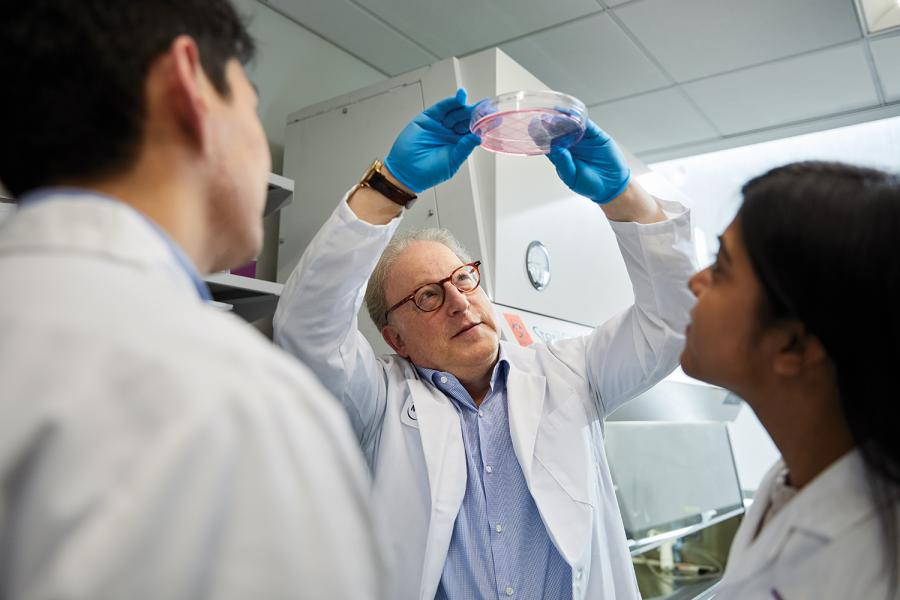
Our laboratory studies the molecular mechanisms involved in the specification of distinct T lymphocyte lineages during development in the thymus and in response to microbial challenge in peripheral tissues. Elucidation of these mechanisms will help us to understand how normal protective immune responses differ from pathogenic ones that result in inflammation and autoimmune disease. Our studies on thymocytes are focused on how they are specified to differentiate towards either the helper or cytotoxic lineages. We are characterizing the transcription factors involved in lineage specification and in regulation of CD4 and CD8 gene expression, with emphasis on mechanisms for establishing epigenetic programs in maturing T cells.
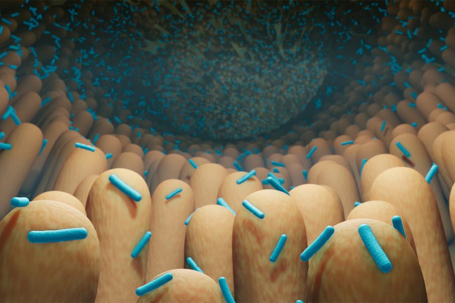
Our studies on peripheral T cells have led to the identification of the nuclear receptor RORgt, that is required for the differentiation of Th17 cells, critical cells in mucosal defense and in inflammatory diseases. We are investigating how distinct commensal microbes influence the differentiation of Th17 cells and other types of T cells, including regulatory T cells (Treg), in different regions of the intestine. A major goal is to determine how intestinal commensal bacteria can trigger systemic T cell-mediated autoimmune diseases, and we are investigating the roles of diverse myeloid cell types, innate lymphoid cells (e.g. ILC3 that are also dependent on RORgt), and cytokines in the polarization of the multiple types of T cells. We are characterizing functions of RORgt in multiple cell types, including ILC3 and mucosal regulatory T cells. We are additionally investigating the communication of microbiota, immune cells, and enteric nervous system cells in intestinal homeostatic functions, e.g. nutrient absorption and regulation of barrier integrity.
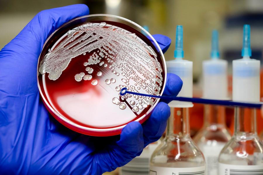
We are seeking to understand how microbial products, e.g. secondary metabolites, influence systemic immune responses to pathogens, tolerance towards normal tissues, and responses to cancer immunotherapy. We also study the potential role of dendritic cells and innate immune system activation in HIV infection to acquire insight into mechanisms of pathogenesis and development of new therapies and vaccines.
212-263-7520
212-263-1498
430 East 29th St
Room 403
New York, NY 10016
Helen L. and Martin S. Kimmel Professor of Molecular Immunology, Department of Pathology at NYU Grossman School of Medicine
MD from Washington University-St Louis
PhD from Washington University-St Louis
Residency, Columbia University, Pathology
Columbia University, Richard Axel
Immunological reviews. 2026 Jan; 337(1):e70082
Nature. 2026 Jan; 649(8096):E1
Nature. 2025 Dec; 648(8092):173-181
Nature. 2025 Jul; 643(8072):776-784
Nature. 2025 Jul; 643(8072):E16
Nature. 2025 Jun; 642(8068):756-765