Main content
Perlmutter Cancer Center
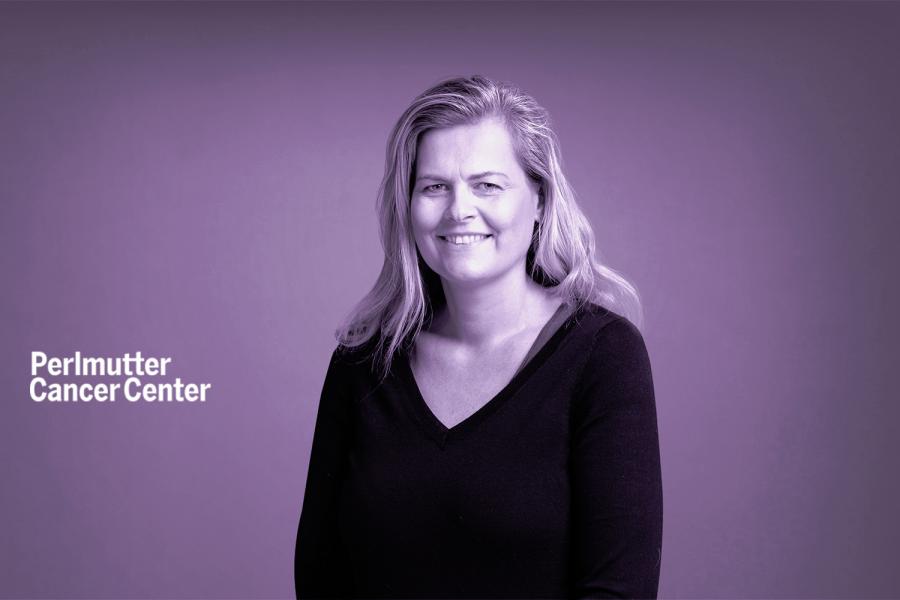
Associate Professor, Department of Pathology
The goal of the Krogsgaard laboratory is to understand the molecular and cellular events that contribute to T-cell sensitivity to ‘self’ (cancer) antigens. To accomplish this we employ advanced and highly innovative methods that combine state-of-the-art biophysical methods for characterizing protein-protein interactions e.g. surface plasmon resonance (SPR), biomembrane force probe X-ray crystallography, infrared (IR) and nuclear magnetic resonance (NMR) spectroscopy with imaging methods (single molecule imaging and FRET) and transgenic TCR technologies which makes our research highly interdisciplinary in its nature. Our research is important not only for understanding the activation of signaling pathways important for T cell function but also for the purpose of modulating signaling through the TCR pharmacologically in new and innovative approaches that can be applied to immunotherapy approaches. This is with the purpose of increasing the sensitivity of T-cells in patients with cancer or HIV or decreasing the sensitivity in patients with autoimmune diseases (multiple sclerosis or diabetes). Supported by the Pew Trust, Cancer Research Institute (CRI), The Arthritis Foundation; New York Chapter, American Cancer Society (ACS), PS-ON/NCI/NIH and NIGMS/NIH.
212-263-9266
Smilow Research Center, 522 First Avenue
7th floor, 701C (office), 707 (lab)
New York, NY 10016
Associate Professor, Department of Pathology at NYU Grossman School of Medicine
PhD from University of Copenhagen
Fellowship, Stanford University, Postdoctoral Fellow
Nature communications. 2023 Jun 23; 14(1):3763
Cell reports. 2016 Mar 29; 14(12):2833-45
Proceedings of the National Academy of Sciences of the United States of America (PNAS). 2013 Apr 23; 110(17):6973-8
Journal of immunology (1950). 2014 Sep 01; 193(5):2587-99CD004345
ACS chemical biology. 2014 Sep 19; 9(9):2165-72
Proceedings of the National Academy of Sciences of the United States of America (PNAS). 2013 Jul 02; 110(27):E2480-9
Annals of clinical & translational neurology. 2022 Oct; 9(10):1643-1659