Main content

Professor, Department of Biochemistry and Molecular Pharmacology
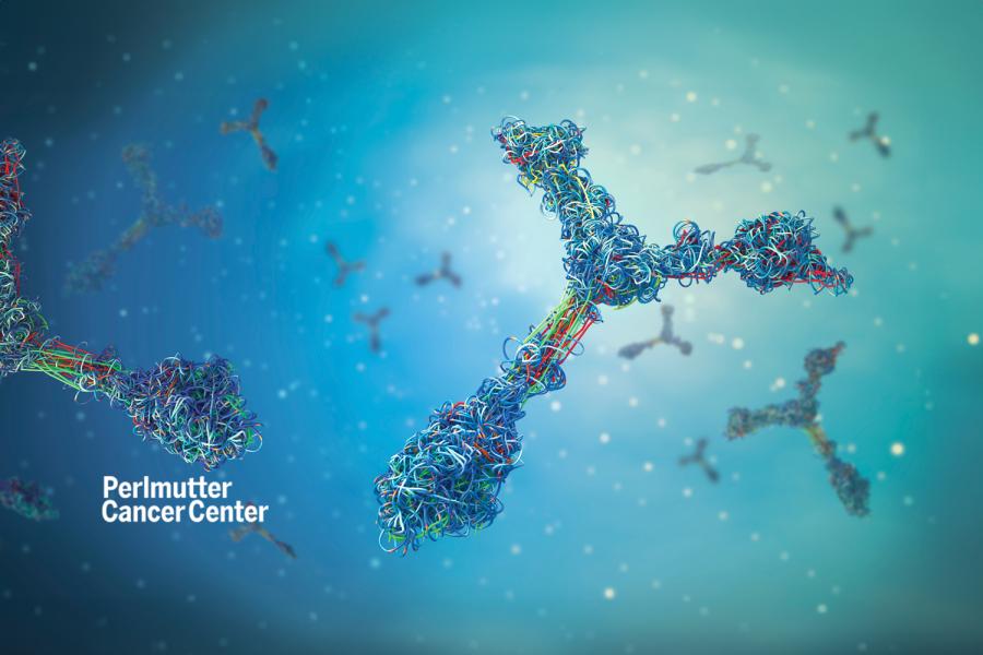
HIV/AIDS is a global pandemic, and the development of a safe and effective HIV vaccine remains one of the most pressing challenges in biomolecular medicine. In a reverse-engineering approach to vaccine discovery, broadly reactive human monoclonal antibodies (mAbs) are identified, and their structures, in complex with their cognate epitopes, are determined. This information is then used to graft the epitopes onto scaffolds to create immunogens that may be capable of eliciting antibodies with a neutralizing breadth similar to that of the broadly reactive mAbs. Therefore, 3D visualization of the HIV epitopes targeted by broadly neutralizing anti-HIV mAbs is crucial for designing immunogens that induce cross-reactive polyclonal antibody responses in mammals.
Researchers at NYU Langone's Kong Lab have collaborated with a team of immunologists, vaccinologists, and computational biologists to characterize a large panel of anti-HIV-1 mAbs, including those targeting the V3, V1V2, and other epitope regions. Our studies of these mAbs not only revealed the structural basis for their broad reactivity but also identified conserved structural elements within the epitope regions that can serve as targets for immunogen design. Consequently, we developed a panel of scaffold immunogens aimed at focusing antibody responses on selected epitope regions and tested them in various animal models, including mice, rabbits, and macaques. Some of these immunogens have been shown to induce potent and durable antibody responses, including broadly neutralizing responses.
212-263-7897
212-263-8166
550 First Avenue, Medical Science Bldg.
3, 398
New York, NY 10016
Course Director, Techniques in Structural Biology & Molecular Biophysics
Leader, Institutional Antibody Production
PhD from Stony Brook University
Frontiers in immunology. 2025 Dec; 16:1722596
Nature communications. 2024 Oct 22; 15(1):9102
Cell death & disease. 2024 Jul 30; 15(7):543
Molecular neurodegeneration. 2024 May 31; 19(1):44
Nature communications. 2024 May 21; 15(1):4301
[Zhong ji yi kan] = [Medicine for intermediate groups]. 2024 Apr 30;
Proceedings of the National Academy of Sciences of the United States of America (PNAS). 2024 Mar 19; 121(12):e2310866121