Main content

Professor, Ronald O. Perelman Department of Dermatology
Professor, Department of Cell Biology
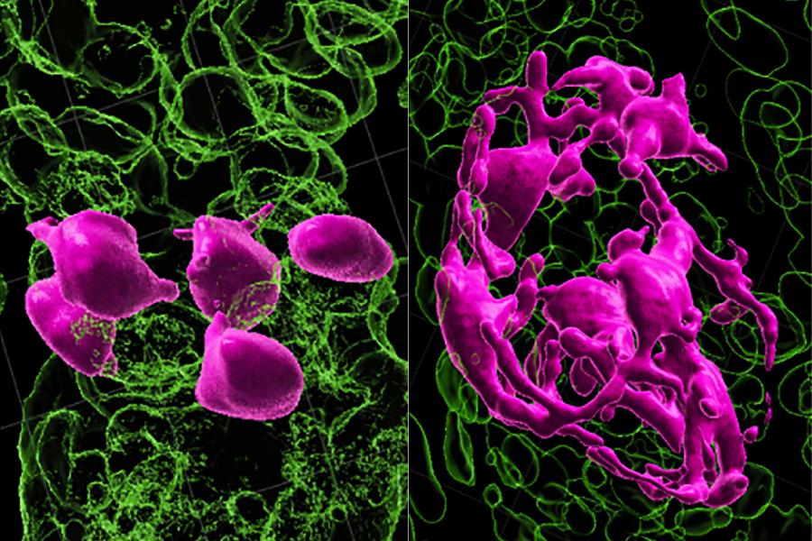
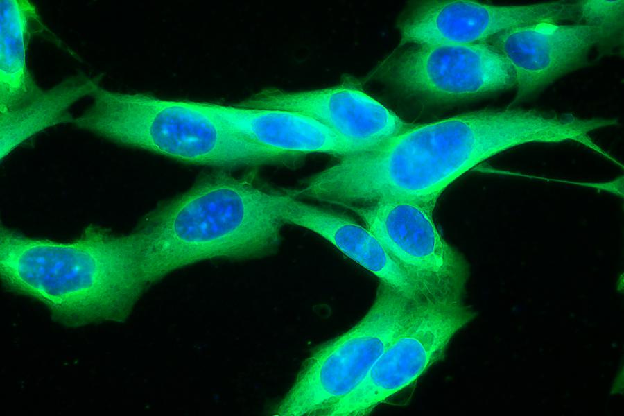
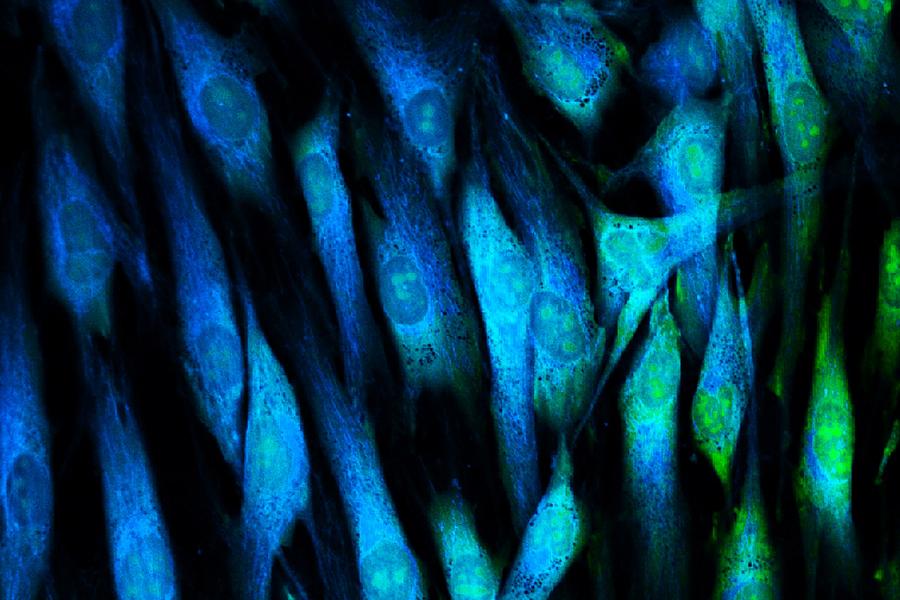
Tissue damage typically results in imperfect repair, calling for research to understand this process. My lab explores several related issues, namely skin wound healing and limb regeneration, to understand why neither results in bona fide tissue regeneration in mammals. In addition, we explore how melanocytes regenerate from stem cells during hair cycling, and we ask how they are dysregulated during aging or in tumorigenesis (i.e. melanoma). For these projects, we have devised unique murine model systems. Our skin wounding model depends upon a novel approach in which new hair follicles can be generated de novo in the healing wound. We manipulate this model to understand how such a regenerative phenomenon can be achieved. Our amputation model takes advantage of the fact that murine distal digit tips can regenerate perfectly whereas more proximal amputations do not undergo regeneration. We examine this disparity to ask why perfect regeneration is achieved in one scenario but not in the other. Our melanocyte model has been developed to track melanocyte stem cells in their progression toward melanocyte regeneration and, in aberrant settings, to hair graying or melanoma. Our projects depend upon genetic manipulation of targeted biological systems, in vivo single cell imaging and single-cell RNA sequencing to address our questions. Through these studies, we aim to understand imperfect repair mechanisms and how they can be manipulated to ultimately achieve perfect tissue regeneration.
212-263-9254
212-263-5819
522 First Avenue
Smilow, 4th Floor, Suite 410
New York, NY 10016
PhD from Nagoya University School of Medicine
Journal of investigative dermatology. 2025 Jan; 145(1):42-49.e2
Developmental biology (Orlando). 2024 May 15; 513:3-11
Journal of investigative dermatology. 2023 Dec; 143(12):2343-2345
Nature. 2023 Apr; 616(7958):774-782
Journal of investigative dermatology. 2022 Oct; 142(10):2565-2569
Nature aging. 2022 Jul; 2(7):568-569
Nature cell biology. 2021 May; 23:439-440
Science advances. 2020 03; 6(12):eaay3704