Main content

Professor, Department of Cell Biology
Professor, Department of Neuroscience
Professor, Department of Psychiatry
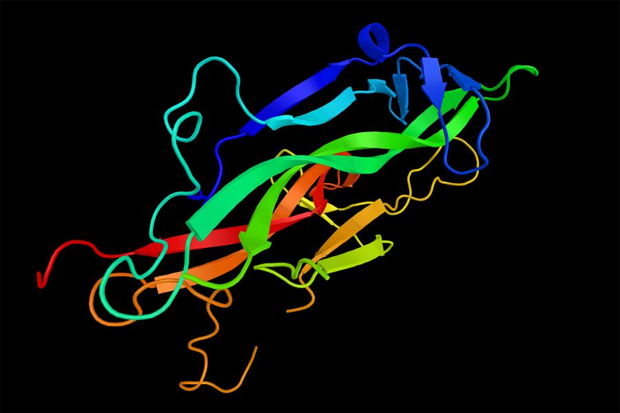
The generation and differentiation of neurons and glial cells are dependent upon cell-cell interactions mediated by a wide variety of growth factors and cytokines. The laboratory is interested in receptor-mediated mechanisms that direct cell differentiation versus cell proliferation in the nervous system. A major goal is to identify the biochemical steps that provide specificity in NGF signaling. Control of cell survival and death by neurotrophins is dictated by an unusual transduction system consisting of two transmembrane receptors, the TrkA tyrosine kinase and the p75 neurotrophin receptor, a member of the TNF receptor superfamily. Members of the NGF family are responsible for neuronal cell survival by activating Trk tyrosine kinases. However, NGF can have the opposite effect, promoting a cell death signal through the p75 receptor. NGF can induce apoptosis of mature oligodendrocytes cultured from rat cerebral cortex. NGF binding to oligodendrocytes expressing the p75 receptor, but not TrkA, resulted in an increase c-jun kinase and caspase activity. Therefore, NGF has the ability of promoting cell survival and cell death in specific cell types through novel signaling mechanisms involving TrkA and p75 receptors. The structural and biochemical features of these two receptors are being defined together with their intracellular signaling mechanisms.
In addition to receptor signal transduction, cell cycle regulation of CNS progenitor cells is being studied. For example, extensive changes in the levels of CDK2 kinase and the cell cycle inhibitor, p27Kip, accompany the differentiation of oligodendrocyte progenitor cells. CDK inhibitors such as p27 negatively regulate G1 phase progression by disrupting cyclin D-CDK4 complexes and cyclin E-CDK2 complexes. The signals necessary for glial cell growth arrest and differentiation are being studied. As a longterm goal, the axonal signals that trigger myelination by oligodendrocyte and Schwann cells will be approached by a combination of molecular and cellular approaches.
212-263-0721
435 East 30th Street, Science Building 11-05
New York, NY 10016
PhD from University of California, Los Angeles
Nature neuroscience. 2025 Nov; 28(11):2296-2309
Cell reports. 2025 Mar 25; 44(3):115344
Proceedings of the National Academy of Sciences of the United States of America (PNAS). 2025 Mar 18; 122(11):e2418249122
Science advances. 2025 Mar 07; 11(10):eadt1763
International journal of eating disorders. 2025 Feb; 58(2):317-335
Cell reports. 2023 Nov 28; 42(11):113333
iScience. 2023 Apr 21; 26(4):106545
Frontiers in molecular neuroscience. 2023 Jul; 16:1179209