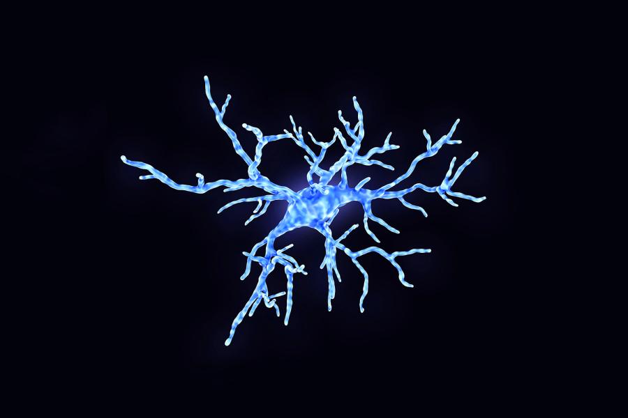
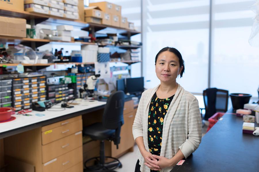
Department of Neuroscience & Physiology

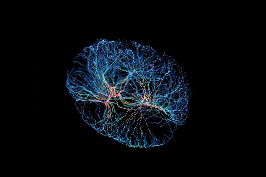
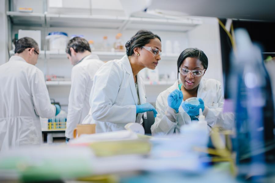
In affiliation with NYU Langone Health’s Neuroscience Institute, the Department of Neuroscience and Physiology is home to an interdisciplinary team of scientists working to enhance our knowledge of fundamental and emerging principles of neuroscience. By bringing together investigators from basic research, translational, and clinical laboratories, we facilitate partnerships that lead to innovative research projects.
Our dynamic educational programs place PhD students, postdoctoral fellows, and research associates in state-of-the-art labs alongside influential faculty scientists.
Through our events series, we connect researchers across NYU Langone and bring neuroscience into our local communities.
Contact Us
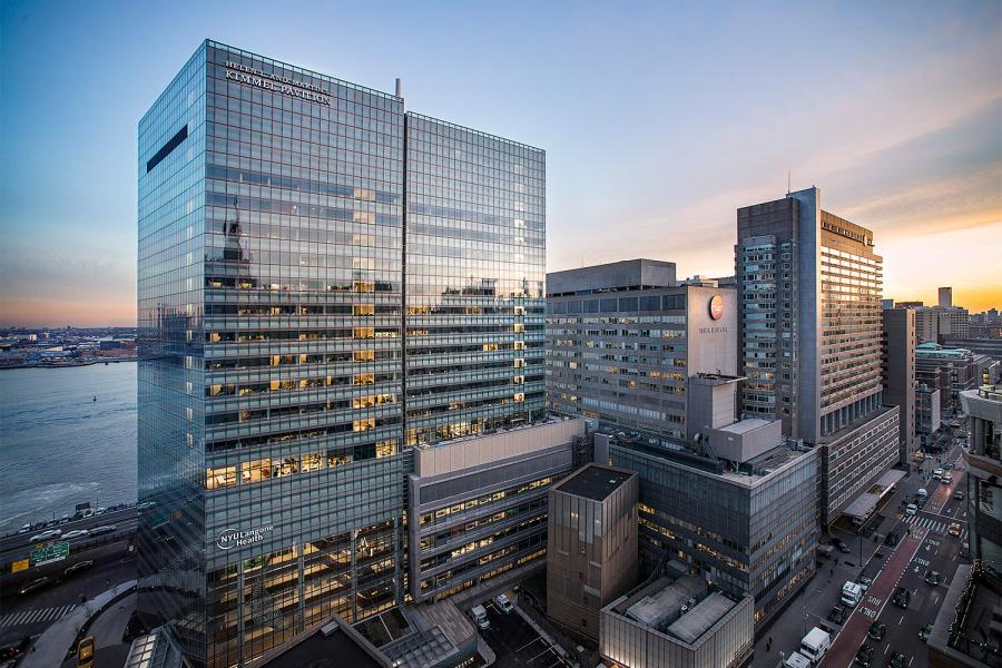
Our offices, laboratories, and conference spaces are located on floors 11 through 13 of NYU Langone’s Science Building, located at 435 East 30th Street in Manhattan. For general inquiries, email us at neuroscience.institute@nyulangone.org.
Our Faculty

Our Events
Graduate Training

Postdoctoral Training

Research Associates

Related News
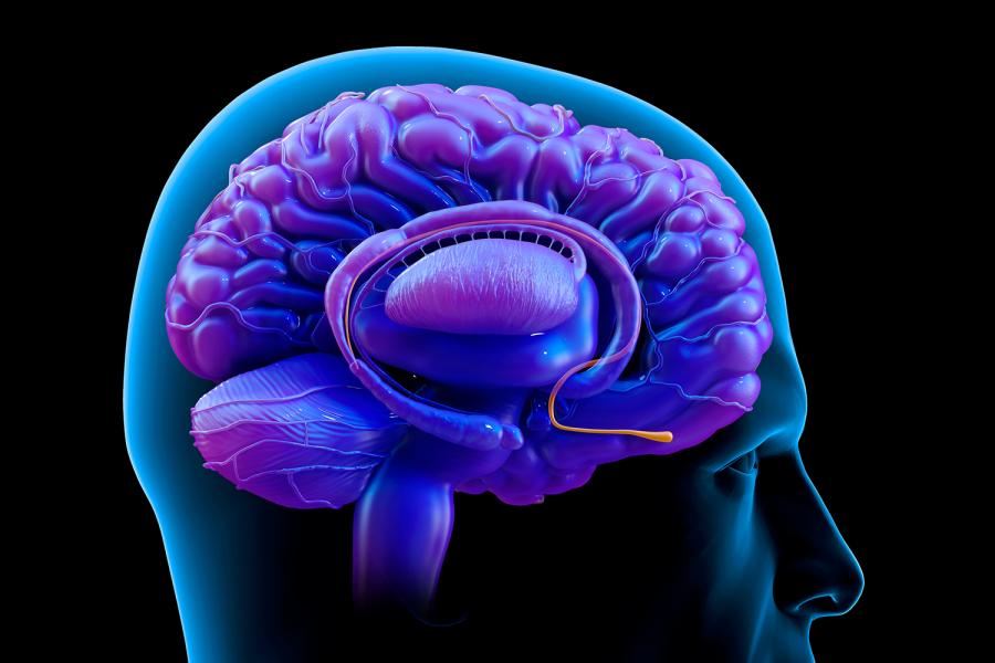
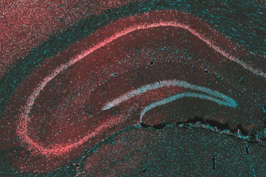
Brain Mechanism Found to Determine Which Memories Last
Brain Mechanism Teaches Mice to Avoid Bullies
Master’s Degree Program in Genome Health Analysis Launches
Brain Circuit Explains Why Infant Cries Prompt Milk Release
‘Ebb & Flow’ Brain Mechanism That Drives Learning Identified
Renowned Researcher Named Director of Neuroscience Institute
Brain Region Prompts Female Mice to Kill Their Young
Restoring Key Brain Rhythm May Help Treat Depression

A $1.2 Million Grant Fuels Research Efforts to Humanize AI
Study Reveals How Cannabidiol Counters Epileptic Seizures
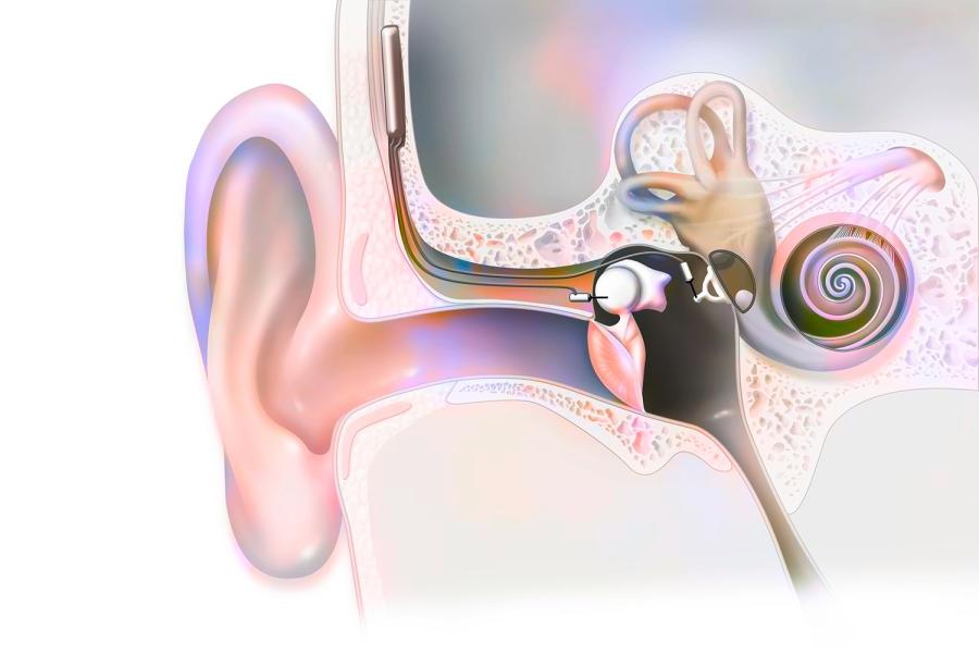
Neuroplasticity May Speed Up Adjustment to Cochlear Implants